
The traditional process of interpreting medical ultrasound images often feels like solving a complex puzzle where clinicians must transform flat two-dimensional slices into a coherent three-dimensional model of the human body. This cognitive transformation, often referred to as mental tomography, demands years of specialized training and places a significant mental burden on healthcare providers during time-sensitive procedures. When a sonographer scans a patient, they are constantly adjusting their spatial understanding to account for the depth and orientation of organs that are only visible as flickering grey planes. This inherent limitation not only slows down the diagnostic process but also introduces a measurable risk of human error, particularly when navigating intricate vascular networks or identifying small lesions. To bridge this gap, researchers have developed Augmented Real-Time Volumetric Imaging in Ultrasound, a groundbreaking system that projects three-dimensional anatomical data directly onto the patient’s skin through an augmented reality headset.
Hardware Integration: The Foundation Of Volumetric Clarity
The Engineering: Designing The Miniature Probe
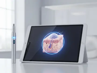
At the heart of this technological leap is a specialized, miniaturized ultrasound probe that measures roughly the size of a standard deck of playing cards, making it highly portable. Traditional volumetric imaging systems are frequently characterized by their bulky frames and exorbitant costs, which often prevent them from becoming standard tools in most clinical settings. This new device utilizes a unique “empty square” array configuration, which strategically positions transducers to capture a vast amount of volumetric data without the need for a dense, high-power sensor grid. By reducing the number of active elements while maintaining high-fidelity output, the engineering team managed to lower both manufacturing costs and power requirements. This design choice ensures that the probe remains cool during extended use and is light enough for a physician to handle with one hand during a procedure. This approach to hardware design prioritizes practicality and ease of use in diverse environments.
Furthermore, the physical dimensions of the probe allow for greater maneuverability compared to the older, more cumbersome 3D scanners currently found in high-end imaging suites. Because the array configuration captures a wider field of view from a smaller footprint, it can easily navigate around the natural contours of the human body, providing a comprehensive look at internal structures. This specific geometry was chosen to optimize the signal-to-noise ratio, ensuring that the resulting digital models are free from the visual artifacts that typically plague low-power ultrasound devices. The transition from traditional linear arrays to this volumetric square format represents a significant shift in how ultrasonic waves are captured and processed. This hardware evolution ensures that the system provides the necessary detail for high-stakes medical decisions while remaining affordable enough for widespread implementation across various healthcare tiers. The result is a device that balances high-end performance with the accessibility of everyday tools.
Software Architecture: Bridging Raw Data And Visualization
To handle the massive stream of information coming from the probe, the system employs a sophisticated chirped data acquisition method that facilitates real-time communication with the headset. This raw data is fed directly into a powerful graphics engine known as Unreal Engine, which is traditionally used to create high-fidelity environments in the gaming industry. The software translates the acoustic reflections into “voxels,” which are essentially the 3D equivalent of pixels, to build a digital twin of the patient’s internal anatomy. By utilizing the massive parallel processing power of modern graphics hardware, the system can update the 3D model in real-time, matching the speed of the physician’s movements. This seamless integration between the acoustic sensors and the rendering engine creates a lag-free experience that is essential for maintaining the illusion of transparency. The software acts as a vital bridge between the physical and digital worlds, ensuring the overlay is precise.
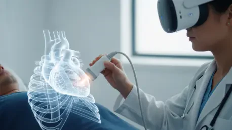
When a physician dons the augmented reality headset, they see a high-resolution representation of the internal organs perfectly aligned with the patient’s physical body. This alignment is achieved through advanced spatial tracking algorithms that ensure the digital model stays locked in place, even if the patient or the physician moves. This sensation of “X-ray vision” allows the user to look through the skin and view the heart, liver, or kidneys as if they were visible to the naked eye. The depth perception provided by the binocular display of the headset is far superior to any 2D monitor, allowing for a natural understanding of how different structures relate to one another in space. This high-fidelity visualization removes the need for the mental reconstruction of anatomy, allowing the clinician to focus entirely on the diagnostic or interventional task at hand. The combination of gaming technology and medical imaging has finally turned the concept of real-time internal visualization into a clinical reality.
Clinical Validation: Measuring Accuracy And User Experience
Empirical Results: Comparing Novices And Experts
To determine the effectiveness of this augmented interface, the development team conducted a series of controlled studies involving a diverse group of medical professionals and novices. Participants were tasked with identifying and locating small objects, such as metallic springs and screws, hidden within tissue-mimicking phantoms. The data revealed that individuals with no prior ultrasound experience were able to perform with a level of accuracy that rivaled seasoned sonographers using traditional 2D equipment. This finding is particularly significant because it suggests that the augmented reality overlay can effectively compress years of specialized training into a more intuitive visual experience. By removing the abstract nature of 2D cross-sections, the system allows anyone with basic medical knowledge to navigate the internal landscape of the human body. The visual feedback provided by the 3D model acted as a powerful guide, reducing the guesswork that often accompanies traditional ultrasound.
Moreover, the study highlighted that the time required to locate a specific target was significantly reduced when using the volumetric overlay. Novices who typically struggled to find depth and orientation on a flat screen were able to reach the target almost immediately once they could see the three-dimensional representation through the headset. While experts still maintained an edge in identifying subtle pathological changes, the gap in spatial localization was nearly eliminated. This equalization of performance demonstrates that the technology is not just an incremental improvement but a fundamental change in how medical imaging can be taught and utilized. The ability for a non-expert to achieve high-precision results opens up new possibilities for emergency care and rural medicine where specialized sonographers may not be available. The empirical evidence supports the idea that making the invisible visible through augmented reality can dramatically improve the baseline of medical performance across the entire industry.
Spatial Navigation: Enhancing The Diagnostic Process
One of the most praised aspects of the new system is the freedom of movement it affords the healthcare provider during an examination. In a traditional setup, the doctor is tethered to a stationary monitor, often forcing them to crane their neck while simultaneously manipulating the ultrasound probe on the patient. With the augmented reality interface, the clinician can lean in, tilt their head, or even walk around the patient to view the internal anatomy from various perspectives. This natural form of navigation provides a level of situational awareness that is impossible to achieve with a fixed 2D display. Many participants in the clinical trials noted a significant reduction in the mental fatigue that usually accompanies long scanning sessions. By aligning the visual data with the physical task, the brain no longer has to work overtime to synchronize hand movements with a separate screen. This ergonomic improvement leads to more thorough examinations and a higher degree of professional comfort.
However, the transition to this new medium was not without its challenges for those who had spent decades mastering traditional 2D ultrasound. Some experts expressed a lingering preference for the older methods, citing the deep-seated muscle memory they had developed over their careers. Despite this initial resistance, these same professionals acknowledged that for high-precision tasks, such as navigating a needle through complex tissue, the 3D spatial context was an undeniable asset. They recognized that the ability to see the target in relation to surrounding blood vessels and nerves provided an extra layer of safety and “peace of mind.” This feedback suggests that while there may be a period of adjustment, the benefits of spatial navigation will eventually outweigh the familiarity of legacy systems. The technology encourages a more interactive approach to diagnostics, where the physician is fully immersed in the patient’s anatomy rather than being a detached observer of a screen. This shift represents a move toward a more intuitive and human-centric medical practice.
Future Implications: From Precision Surgery To Global Health
Clinical Applications: Improving Interventional Success
The practical utility of this volumetric imaging system extends far beyond simple diagnostic scans and into the realm of complex interventional procedures. For example, during a guided needle biopsy, a physician must navigate a small needle toward a specific lesion while avoiding vital structures like arteries and nerves. By seeing a 3D model of the target directly under the skin, the practitioner can plan the trajectory with absolute precision, ensuring the needle reaches its destination on the first attempt. This level of accuracy minimizes trauma to the surrounding tissue and significantly reduces the risk of post-procedural complications. In addition to biopsies, the system is exceptionally well-suited for monitoring dynamic organs such as the heart. Tracking the rapid movements of heart walls and valves in real-time provides critical feedback during echocardiographies, where understanding the spatial relationship of moving parts is vital for diagnosing structural heart disease.
Furthermore, the real-time nature of the system allows for the immediate adjustment of surgical plans if the internal conditions change during a procedure. If a target shifts due to the patient’s breathing or the introduction of a surgical tool, the digital twin updates instantly to reflect the new reality. This dynamic feedback loop ensures that the physician is always working with the most current information, rather than relying on static images taken before the procedure began. The potential for integrating this technology with robotic-assisted surgery is also substantial, as it could provide the robotic system with a precise roadmap of the patient’s unique anatomy. By providing a clear, uninterrupted view of the surgical field beneath the surface, the augmented reality interface acts as a safety net that enhances the skill of the surgeon. The focus on interventional accuracy ensures that patients receive the most effective care possible with the least amount of invasive interference. This evolution in surgical guidance marks a major step toward a future of zero-error medical procedures.
Healthcare Evolution: Accessibility And Education
The long-term impact of augmented volumetric imaging will likely be felt most strongly in the areas of medical education and global healthcare accessibility. Because the system utilizes relatively low-power and inexpensive hardware, it is ideally suited for deployment in resource-limited regions where high-end medical imaging is currently unavailable. In these settings, the intuitive nature of the AR-VIU interface could allow local healthcare workers to perform essential diagnostic scans with minimal training, potentially saving countless lives through earlier detection of diseases. Additionally, the system could be used for remote monitoring, where data from a home-based probe is streamed to a specialist in another city for a real-time consultation. This democratization of specialized imaging has the potential to bridge the gap in healthcare quality between urban centers and rural communities. By making high-quality diagnostics portable and easy to understand, the technology empowers a broader range of providers to deliver expert-level care.
In the classroom, this technology had already begun to revolutionize how medical students learn human anatomy and diagnostic techniques. Instead of studying static diagrams or cadavers, students can interact with living, breathing 3D models that respond to their movements. The development team successfully demonstrated that this interactive approach drastically shortened the time required to master ultrasound techniques, as the direct visual feedback reinforced the relationship between hand placement and internal views. The next steps for the research focused on increasing image resolution and shrinking the hardware even further to facilitate a wider range of clinical applications. These advancements paved the way for the standardization of “X-ray vision” as a common tool in every hospital, clinic, and medical school. As the technology matured, it moved closer to becoming an indispensable part of the modern medical toolkit, ensuring that the internal mysteries of the human body are no longer hidden behind a screen. The era of intuitive, spatial medicine was finally within reach for everyone.