
The realization that a routine heart scan holds the hidden blueprint of our metabolic longevity has fundamentally altered the landscape of preventative diagnostic imaging. Medical professionals are no longer limited to examining the plumbing of the heart but are now looking at the structural integrity of the surrounding tissue to predict future outcomes. This evolution represents a significant leap forward in how technology bridges the gap between static imagery and dynamic health forecasting. By analyzing what was once considered secondary data, clinicians can now provide a more nuanced picture of a patient’s survival probability and cardiovascular health.
This technology introduces an automated method for evaluating skeletal muscle health by analyzing routine diagnostic scans. Traditionally, imaging like coronary computed tomography angiograms focused strictly on arterial blockages to determine a patient’s immediate risk of cardiac events. However, AI-driven analysis now allows clinicians to assess muscle attenuation, which measures the density and composition of the muscle tissue to identify intramuscular fat marbling. This shift marks a transition toward holistic diagnostic profiling, where skeletal muscle serves as a vital indicator of internal cardiovascular resilience and overall longevity.
Introduction to AI-Driven Muscle Quality Assessment
Instead of relying on basic body mass index or weight measurements, this approach prioritizes the qualitative state of the tissue. It recognizes that two individuals with identical weights may have vastly different internal health profiles based on their muscle-to-fat ratios. This nuanced understanding allows for more personalized healthcare, moving away from a one-size-fits-all model. By leveraging existing scans, the technology adds value to medical procedures that have already been performed, making the healthcare process more efficient and data-rich.
Furthermore, this diagnostic shift emphasizes that muscle is not merely a tool for movement but a metabolic organ. The presence of fat within the muscle fibers often correlates with systemic inflammation and poor metabolic health. By identifying these issues early through routine imaging, medical providers can address the root causes of frailty before they manifest as chronic diseases. This proactive approach is essential for an aging population that requires more sophisticated monitoring of physical integrity.
Core Features and Technical Capabilities
Automated Skeletal Muscle Attenuation Measurement
The primary function of this technology is the measurement of muscle brightness or attenuation on a digital scan. High-quality muscle tissue with minimal fat infiltration appears lighter and denser because of how X-ray beams interact with the tissue. The AI distinguishes between sheer muscle mass and tissue quality, providing a more accurate health metric than size alone. This allows for the identification of fat marbling in humans, which has become a significant marker for poor cardiovascular outcomes in recent clinical studies.
This precision is what differentiates this tool from standard volumetric measurements that might overestimate the health of a sedentary person with large but fatty muscles. By focusing on density, the software provides a clear metric that reflects the functional capacity of the muscle. This objective data helps clinicians steer patients away from general weight loss and toward specific goals like improving muscle quality through resistance training and metabolic optimization.
Rapid Machine Learning Diagnostic Processing
A standout performance characteristic of this technology is its processing speed. While a human radiologist might require several hours to manually segment and measure muscle, bone, and fat across a single scan, the AI algorithm completes these complex calculations in under sixty seconds. This efficiency enables the extraction of deep health insights from existing diagnostic imagery without requiring additional procedures or significant time investments from medical staff.
Moreover, the speed of processing allows for the large-scale analysis of patient populations, which was previously impossible. When diagnostic results are available almost instantly, they can be discussed during the same consultation where the heart scan results are reviewed. This immediate feedback loop is critical for patient engagement, as it connects the abstract data of a scan to actionable lifestyle changes while the patient is still in the clinic.
Emerging Trends in Diagnostic Imaging
The field is moving toward opportunistic screening, where AI extracts secondary health data from scans performed for other purposes. Current trends show a shift away from isolated metrics, such as calcium scoring, toward a comprehensive assessment of the patient’s physical frailty and metabolic health. Industry behavior is increasingly favoring these multi-faceted AI tools to provide a more complete picture of patient risk during standard medical checkups from 2026 to 2028.
In addition to cardiovascular health, these trends are influencing how oncology and geriatrics approach patient care. By understanding the muscular baseline of a patient, doctors can better predict how well an individual will tolerate aggressive treatments like chemotherapy or major surgery. The ability to pull this data from a standard CT scan without extra radiation exposure makes it a highly sustainable trend in modern clinical practice.
Real-World Applications and Use Cases
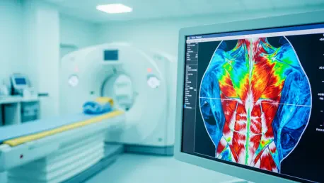
In clinical settings, AI muscle analysis is being deployed to refine cardiovascular risk stratification. For example, patients undergoing routine heart scans can now receive a concurrent assessment of their torso and back muscles, such as the erector spinae and pectorals. This data is used to predict the ten-year risk of heart attack and mortality, with high-quality muscle correlating to a 31 percent reduction in heart attack risk. These findings are prompting healthcare providers to prescribe specific physical interventions.
Patients with low muscle density are being directed toward functional strength training or programs like pilates that specifically target the core and intercostal muscles. These prescriptions are no longer generic advice but are backed by objective imaging data that shows exactly where the patient is lacking. This application of the technology turns a diagnostic tool into a motivational one, providing patients with a clear “before and after” metric for their physical improvement.
Technical Hurdles and Implementation Challenges
Despite its promise, the technology faces obstacles regarding the standardization of imaging data across different CT scanner manufacturers. Variations in X-ray intensity and image resolution can affect attenuation readings, requiring more robust calibration to ensure consistency. Integrating these AI tools into the high-pressure environment of radiology departments also requires seamless software interoperability to ensure the data is used safely for clinical decision-making.
Furthermore, there is the challenge of regulatory approval and the development of universal benchmarks for what constitutes healthy muscle density across different age groups. Without these standards, interpreting the data remains a complex task for general practitioners. Overcoming these hurdles will require collaboration between AI developers and medical device manufacturers to create a unified ecosystem that prioritizes data accuracy over proprietary software constraints.
Future Outlook and Long-Term Impact
The trajectory of AI muscle density analysis points toward a more unified view of physical fitness and internal health. Future developments are expected to expand this analysis to other muscle groups and integrate it with longitudinal health data to monitor the effectiveness of exercise programs in real-time. Long-term, this technology could become a standard component of preventative medicine, fundamentally changing how we define fitness by prioritizing tissue quality over physical appearance.
As the software becomes more accessible, it may even move into the consumer space through lower-intensity scanning or integration with other wearable health monitors. The ultimate goal is to create a continuous feedback loop of physical health that allows individuals to maintain their vitality as they age. This will likely reduce the burden on healthcare systems by preventing the onset of frailty-related complications and chronic metabolic disorders.
Summary of Clinical Findings and Assessment
The review of AI muscle density analysis indicated a major breakthrough in understanding the link between skeletal integrity and cardiovascular health. By providing a rapid, objective measure of muscle quality, this technology offered a powerful new tool for identifying at-risk individuals who might otherwise have been overlooked by traditional screenings. The clinical data demonstrated that the density of torso muscles served as a reliable predictor for long-term mortality, independent of existing heart disease markers.
The evaluation suggested that the next logical step involved the widespread integration of these AI modules into existing radiology workflows. Stakeholders moved toward establishing clearer clinical guidelines to translate muscle attenuation scores into specific exercise and nutritional interventions. These actionable insights ensured that the technology did more than just provide data; it facilitated a cultural shift in medicine toward the preservation of functional strength. Ultimately, the successful deployment of these tools confirmed that the future of longevity rested in a more comprehensive understanding of the body’s structural health.