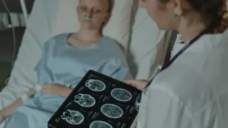

Introduction
The devastating impact of central nervous system tumors on pediatric health continues to drive a relentless search for more effective therapeutic strategies that can offer hope to families facing a grim diagnosis. For decades, the complexity of the human brain and the aggressive nature of childhood cancers have made it difficult to develop treatments that are both safe and effective. While medical science has made significant strides, the primary obstacle has remained a lack of accessible and accurate laboratory tools that can truly simulate how a human tumor behaves within a living patient.
This article explores the pioneering work at St. Jude Children’s Research Hospital, where scientists have successfully developed sophisticated 3D organoid models to study some of the most lethal pediatric brain tumors. The objective is to explain how these new models function, why they represent a significant improvement over previous methods, and what their implementation means for the future of personalized medicine. Readers will gain insight into the biological validation of these models and the collaborative spirit driving this open-science initiative.
Key Questions or Key Topics Section
Why are Traditional Research Models Insufficient for Pediatric Brain Cancer?
The traditional gold standard for cancer research has long been the patient-derived orthotopic xenograft, a method involving the transplantation of human tumor cells into animal models. While these models are effective at retaining many of the tumor’s primary characteristics, they are inherently limited by several practical factors. The establishment of such models is a laborious process that can take many months, which is a timeframe that rarely aligns with the clinical urgency required for treating aggressive pediatric embryonal tumors.
Furthermore, the high costs and specialized infrastructure needed to maintain these animal-based systems often restrict their use to the most well-funded research centers. This centralization of resources creates a disparity in research capabilities, preventing smaller labs from contributing to the development of new therapies. In contrast, the pursuit of more agile and cost-effective modeling techniques is essential to democratizing pediatric oncology research and ensuring that scientific breakthroughs are not stalled by logistical or financial barriers.
How do 3D Organoids Replicate the Complexity of Human Brain Tumors?
The breakthrough at St. Jude involves the creation of patient-derived 3D tumor organoids, which are simplified, lab-grown versions of human organs or tumors. Unlike traditional 2D cell cultures that grow flat on a plastic dish, 3D organoids allow cells to organize into complex structures that better mimic the environment of a human brain. This structural complexity is vital because the way cancer cells interact with their neighbors and their physical surroundings influences how they grow and how they respond to chemotherapy.
By focusing on aggressive malignancies like medulloblastoma and atypical teratoid rhabdoid tumors, researchers have successfully engineered environments where these cells thrive outside the human body. These organoids can also be used to create tumor organoid xenografts, providing a hybrid model that allows for more detailed observation of tumor behavior in a living system. This dual approach provides a powerful platform for testing how different cancers spread and identify the unique vulnerabilities of each specific tumor type.
What Methods Ensure the Biological Accuracy of These New Models?
A significant challenge in creating any laboratory model is ensuring that it remains a faithful representation of the original human disease over time. To prevent biological drift—a phenomenon where the lab-grown cells lose their original characteristics—the research team at St. Jude utilized a comprehensive suite of molecular profiling tools. This process involved comparing the genetic and epigenetic signatures of the organoids directly with the patient samples from which they were derived.
The scientists employed advanced techniques such as DNA methylation profiling and whole-genome sequencing to verify that the genetic blueprint of the cancer remained intact. Additionally, single-cell RNA sequencing was used to examine the individual cells within the organoids, confirming that the cellular diversity found in a patient’s tumor was accurately reflected in the lab. This rigorous validation ensures that when a drug shows promise in the organoid model, there is a high degree of confidence that it will behave similarly in a clinical setting.
How do These Advancements Accelerate the Search for New Treatments?
The primary benefit of these 3D models is the speed and efficiency they bring to the drug discovery process. Because organoids can be grown and expanded relatively quickly in a laboratory setting, they allow for high-throughput screening of hundreds of different therapeutic compounds. This rapid testing capability means that researchers can identify potential treatments in a fraction of the time it would take using traditional animal models alone, potentially moving life-saving drugs toward clinical trials much faster.
Moreover, the hospital’s decision to share these validated models with the global scientific community marks a significant shift toward open-source medical research. By removing the need for every laboratory to procure their own fresh patient samples, which are rare and difficult to obtain, St. Jude is providing a standardized resource that can be used by scientists worldwide. This collaborative framework encourages a more unified effort to solve the complexities of pediatric brain cancer, ensuring that the collective knowledge of the global scientific community is leveraged to find a cure.
Summary or Recap
The development of patient-derived 3D organoids at St. Jude Children’s Research Hospital represents a transformative shift in the field of pediatric neuro-oncology. These models offer a unique combination of biological accuracy and operational efficiency, overcoming the significant time and cost barriers associated with previous research methods. By utilizing rigorous genetic and epigenetic validation, the team has ensured that these lab-grown tumors serve as reliable proxies for human disease, providing a stable platform for therapeutic testing.
Key takeaways include the scalability of these models for drug screening and the democratization of research through the sharing of resources with the international community. This approach not only streamlines the path from the laboratory to the patient’s bedside but also fosters a collaborative environment where rare diseases can be studied with greater intensity. As these tools become more widely integrated into oncology, the potential for discovering targeted, less toxic treatments for children increases significantly, highlighting the importance of continued innovation in tissue engineering and molecular biology.
Conclusion or Final Thoughts
The successful creation and validation of these advanced 3D models demonstrated that the gap between laboratory research and clinical application could be narrowed through innovative bioengineering. This initiative moved beyond mere scientific curiosity and addressed the urgent practical needs of the pediatric oncology community. By establishing a faster and more accessible system for studying aggressive tumors, the research provided a foundation for future breakthroughs that were previously hindered by logistical constraints.
Researchers and clinicians were encouraged to adopt these standardized models to harmonize their efforts and improve the reproducibility of their findings. The shift toward a more collaborative and open-science model suggested that the next generation of cancer treatments would be born from shared knowledge rather than isolated discoveries. Ultimately, this work reflected a deep commitment to changing the trajectory of pediatric brain cancer, ensuring that the progress made in the lab translated into meaningful improvements in patient survival and long-term health outcomes.


