
Recent research suggests that the prognostic value of the CFH gene may be driven primarily by the FHL-1 isoform rather than the circulating full-length protein. In the rapidly shifting landscape of 2026 oncology, identifying reliable prognostic biomarkers has become the cornerstone of improving patient survival and refining therapeutic strategies. The CFH gene, which encodes complement factor H, has recently emerged as a focal point in expansive pan-cancer analyses. This gene acts as a vital regulator of the complement system, a key component of the innate immune response responsible for identifying pathogens and clearing cellular debris. Across various tumor types, high levels of CFH expression consistently correlate with advanced clinical stages, poor patient survival, and increased immune cell infiltration. However, recent critiques suggest that the current enthusiasm for CFH as a universal biomarker may be mathematically imprecise due to a fundamental measurement oversight. The biological reality is that this single gene produces two distinct protein products that are often lumped together in data sets, potentially obscuring the true driver of cancer progression.
The Structural Divide: Understanding FH and FHL-1
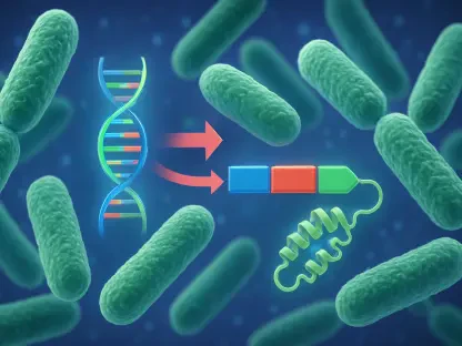
The complexity of the CFH gene is rooted in its ability to produce two entirely different proteins through the process of alternative splicing. Although they share a common genetic origin, full-length complement factor H (FH) and factor H–like 1 (FHL-1) possess radically different physical structures that dictate their behavior in the body. FH is a massive glycoprotein consisting of twenty complement control protein domains that circulates primarily in the blood plasma. Its primary role is to act as a sentinel, distinguishing healthy host cells from foreign threats and preventing the uncontrolled activation of the immune system. Because of its large size and extensive sugar coatings, FH is well-suited for systemic regulation but faces physical limitations when attempting to move from the bloodstream into the dense, pressurized environments of solid tumors. This structural mass makes FH a stable presence in the circulation but a potentially less active participant within the internal architecture of a malignant growth.
In contrast, FHL-1 is a much smaller isoform that contains only the first seven domains of the full-length protein, ending with a unique C-terminal tail not found in the larger molecule. While FHL-1 retains the core machinery necessary for immune regulation, it lacks the specific recognition domains that anchor FH to various host surfaces. This structural streamlined nature allows FHL-1 to perform functions that its larger counterpart cannot easily duplicate. The divergence between these two proteins means that although they originate from the same genetic blueprint, they effectively operate as two different tools with distinct geographical targets. By treating them as a single entity in genomic studies, researchers risk missing the specific nuances of how each protein contributes to the tumor microenvironment. Understanding this structural divide is the first step toward untangling the conflicting data often seen in modern cancer research where CFH expression is used as a broad catch-all for immune activity.
Spatial Dynamics: Mobility within the Tumor Microenvironment
A major functional distinction between FH and FHL-1 lies in their relative ability to navigate the complex physical barriers of the tumor microenvironment. FHL-1, being significantly smaller and lacking the heavy glycosylation of FH, possesses a superior capacity to diffuse through dense biological tissues. This mobility is particularly critical when considering the extracellular matrix (ECM), the thick network of proteins that surrounds solid tumors and often acts as a shield against immune cells and therapies. Research in specialized fields, such as ophthalmology and neuro-oncology, has already demonstrated that FHL-1 is the dominant regulator in matrix-rich environments like the eye’s Bruch’s membrane or within the aggressive boundaries of glioblastoma. In these contexts, the smaller isoform can penetrate deep into the tissue to regulate local immune responses, whereas the larger FH molecule remains restricted to the peripheral vasculature or the immediate surface of the tumor.
Furthermore, evidence suggests that FHL-1 is not just a passive migrant from the blood but is often produced directly by the cancer cells themselves. In malignancies of the lungs and pancreas, cancer cells have been observed secreting FHL-1 into their immediate surroundings, effectively “bio-hacking” their own microenvironment to suppress local immune attacks. This localized production creates a high concentration of the small isoform exactly where the tumor is most vulnerable to immune infiltration. Because FHL-1 can anchor itself to the matrix more effectively than the circulating FH, it becomes a permanent resident of the tumor site. This spatial dynamic implies that when researchers measure CFH activity within a tumor biopsy, they are likely observing the localized effects of FHL-1 rather than the systemic influence of FH. Distinguishing between these two spatial roles is essential for understanding how a tumor successfully evades the host’s innate immune defenses.
Methodological Risks: The Dangers of Aggregate Genomic Signals
The technical foundation of many current pan-cancer studies relies on quantitative reverse-transcription PCR (qRT-PCR) to measure gene expression levels, but this method carries a significant risk of signal contamination. Most common primers used in these assays target the N-terminal region of the CFH gene, which is a sequence shared by both the FH and FHL-1 transcripts. As a result, the data produced by these studies represents an “aggregate signal” that combines the expression of both proteins into a single numerical value. This lack of resolution makes it impossible to determine whether a high CFH score is driven by the circulating FH or the localized FHL-1. If one isoform is a marker of aggressive disease while the other is merely a bystander, the current data remains fundamentally “noisy.” This ambiguity complicates the interpretation of how the complement system actually interacts with cancer, leading to potential misidentifications of the primary biological actor.
This measurement problem also extends to functional experiments involving gene silencing or “knockdowns.” When researchers use tools like short hairpin RNA (shRNA) to suppress CFH expression to observe changes in cell behavior, they frequently target these same shared N-terminal sequences. Consequently, both FH and FHL-1 are eliminated from the system simultaneously. While this may result in observable changes in cell migration or proliferation, it leaves a critical question unanswered: which specific protein loss caused the effect? It is entirely possible that the observed reduction in tumor growth is due solely to the loss of FHL-1, but the current experimental designs would attribute the result to the entire CFH gene family. Without isoform-specific interventions, the scientific community continues to operate with a blurred vision of the underlying molecular mechanisms, which can lead to the development of therapies that are poorly targeted or less effective than they could be.
Clinical Hurdles: Translating Molecular Data into Diagnostics
The transition from lab discovery to a validated clinical diagnostic tool requires a level of precision that aggregate signals cannot provide. In the current era of precision medicine, clinicians depend on specific molecular signatures to determine the best course of treatment for an individual patient. If a diagnostic test cannot distinguish between FH and FHL-1, its accuracy may vary significantly depending on the type of sample being analyzed. A blood-based liquid biopsy will naturally be dominated by the high concentrations of circulating FH, while a solid tissue biopsy will likely reflect the matrix-retained FHL-1. If the true driver of a patient’s prognosis is the localized FHL-1, a blood test focusing on the broader CFH signal might provide a misleading picture of the disease’s progression. This discrepancy could lead to inconsistent results across different clinical settings, undermining the reliability of the biomarker.
Moreover, the lack of isoform resolution could result in suboptimal treatment decisions for patients with complex or late-stage cancers. For instance, if a correlation between CFH and survival is driven entirely by FHL-1 in pancreatic cancer but by FH in a different malignancy, a universal “CFH-high” diagnostic criteria would fail to account for these tissue-specific differences. This could result in patients receiving immunotherapies or complement-targeting drugs that are not actually suited to the specific molecular environment of their tumor. To move forward, diagnostic development must shift away from general gene expression and toward isoform-specific detection. By isolating the signal of the most relevant protein, clinicians can better predict how a tumor will respond to specific interventions, ultimately leading to more personalized and effective care. The ability to differentiate between these two molecules is not just a technical detail; it is a clinical necessity for the future of oncology.
Technological Solutions: Moving Toward Isoform Specificity
Overcoming the measurement problem requires the widespread adoption of advanced analytical technologies that can resolve the specific transcripts of the CFH gene. Splice-junction specific PCR is one such method, allowing researchers to design primers that target the unique connection points found only in the FHL-1 isoform or the distal regions of FH. This approach provides a clear separation of data, ensuring that the expression levels of each protein are tracked independently. Additionally, long-read RNA sequencing has become an invaluable tool in 2026, as it allows for the sequencing of entire transcript molecules in a single pass. This technology eliminates the guesswork associated with piecing together short genetic fragments, making it easy to identify exactly which splice variants are present in a given sample. By integrating these methods into standard research workflows, the scientific community can begin to correct the historical biases found in earlier pan-cancer data sets.
Beyond the transcript level, advanced proteomics and mass spectrometry offer a way to confirm these findings at the protein level. These techniques can distinguish FH from FHL-1 based on their differing molecular weights and unique peptide sequences, providing a definitive map of protein distribution within the tumor. This level of molecular detail is essential for verifying that the transcript levels measured in the lab actually translate to functional proteins within the patient. As these sophisticated tools become more accessible, they will allow for a “constructive refinement” of previous research, building a more accurate foundation for the next generation of biomarkers. The path toward better cancer prognostics lies in embracing this molecular complexity rather than oversimplifying it. By focusing on the unique roles of FH and FHL-1, researchers can unlock a much clearer understanding of the complement system’s role in cancer, paving the way for more precise and impactful diagnostic tools.
The Importance of Molecular Specificity
The investigation into the CFH gene provided a broader lesson for the entire field of transcriptomics and genomic research. While large-scale “big data” projects offered incredible statistical power, their ultimate utility was limited by the specificity of the tools used to collect the information. This study demonstrated that genes undergoing alternative splicing represent a hidden layer of complexity that can easily lead researchers toward incorrect conclusions if not properly accounted for. The discrepancy between FH and FHL-1 served as a high-stakes reminder that the most significant biological drivers often reside in the details of isoform variation. Moving forward, the scientific community has begun to prioritize the development of “isoform-aware” experimental designs to ensure that the signals detected in the lab are truly representative of the underlying pathology.
To ensure the successful translation of these findings into clinical practice, several actionable steps were identified for future research. Scientists were encouraged to re-examine existing pan-cancer databases using newer, more specific bioinformatic pipelines that can differentiate between splice variants. Furthermore, the development of monoclonal antibodies that specifically target the unique C-terminal tail of FHL-1 became a priority for creating more accurate immunohistochemistry assays. By refining the measurement of these two proteins, the field of oncology moved closer to a future where biomarkers are not just statistical correlations but precise mirrors of tumor biology. The transition toward molecular specificity has proven to be the most effective way to harness the potential of the complement system, transforming it from a source of noisy data into a reliable tool for saving lives. Final evaluations of this genomic era highlighted that the difference between a promising lead and a successful clinical tool often depended on the ability to distinguish seven protein domains from twenty.