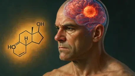

The medical community has long perceived male sex hormones as catalysts for various malignancies, yet recent findings regarding glioblastoma indicate that these same biological components might actually serve as vital protectors for the human brain. Glioblastoma multiforme remains one of the most lethal and aggressive forms of brain cancer, disproportionately affecting men with both greater frequency and significantly shorter survival rates. Historically, the oncological consensus held that androgens like testosterone were strictly detrimental factors, as they are well-documented to suppress immune responses in cancers of the lung, bladder, and skin. However, researchers at the Cleveland Clinic have published a groundbreaking study in the journal Nature that fundamentally challenges this long-held assumption. Their work suggests that within the unique environment of the central nervous system, testosterone acts as a crucial regulatory agent rather than a promoter of disease. By maintaining the functional stability of the brain’s immune landscape, this hormone prevents a systemic collapse of the body’s defense mechanisms. This shift in perspective marks a significant milestone in cancer neuroscience, demonstrating that the anatomical location of a tumor dictates how the endocrine system interacts with the immune response.
The Mechanism: How Hormonal Shifts Impact Brain Immunity
Central to this discovery is the intricate relationship between testosterone and microglia, which are the specialized immune cells tasked with patrolling the brain environment. Under normal physiological conditions, testosterone serves to regulate these cells, keeping their inflammatory signaling in check to prevent overactivity. When testosterone levels are suppressed—either through natural aging or medical intervention—these microglia begin to malfunction and trigger a localized inflammatory response. This inflammation is not confined to the brain; it effectively signals the hypothalamic-pituitary-adrenal axis, which is the body’s primary stress-response system. Once activated by this specific brain inflammation, the HPA axis releases a cascade of stress hormones that travel throughout the entire body. This systemic reaction ultimately leads to a broad suppression of the immune system, particularly affecting the efficacy of cancer-fighting T-cells that are necessary to identify and destroy malignant cells within the nervous system. The identification of this chain reaction provides a clear biological explanation for why traditional androgen-blocking therapies have failed to benefit brain cancer patients.
This biological chain reaction highlights a profound paradox where the absence of testosterone creates a more hostile environment for the patient than its presence. In typical solid tumors found outside the brain, high androgen levels are often associated with reduced T-cell activity, leading clinicians to utilize androgen deprivation therapy as a standard treatment. In contrast, the Cleveland Clinic study demonstrates that the brain possesses a unique regulatory feedback loop where testosterone is required to prevent the HPA axis from overreacting. Without this hormonal buffer, the resulting stress response creates a state of systemic exhaustion for the immune system, leaving the body unable to mount an effective defense against glioblastoma. This research underscores the necessity of considering the specific neural and endocrine context of a disease rather than applying broad oncological rules across all tumor types. Such findings suggest that treating glioblastoma requires a complete reversal of the strategies used for other cancers, focusing on hormonal preservation rather than suppression to maintain immune integrity.
Clinical Correlations: Translating Laboratory Insights into Patient Outcomes
The researchers successfully synthesized these preclinical observations with extensive human clinical data to provide a comprehensive view of how testosterone levels influence patient longevity. They observed a distinct trend in cancer registry data showing that male T-cell counts naturally decline with age, a phenomenon that correlates directly with the gradual reduction of testosterone levels in the aging male population. This biological decline often mirrors the peak onset of glioblastoma, suggesting that the loss of hormonal protection may be a contributing factor to the increased vulnerability seen in older men. Furthermore, an analysis of historical patient records revealed a significant real-world correlation: male glioblastoma patients who received supplemental testosterone therapy alongside their standard chemotherapy lived longer on average than those who did not receive such support. This finding provides compelling evidence that maintaining or restoring physiological testosterone levels can improve the durability of the immune system, thereby enhancing the body’s ability to combat the aggressive progression of brain tumors over an extended period.
The study shifted the paradigm of glioblastoma treatment by establishing testosterone as a potential therapeutic ally rather than a risk factor. Moving forward, the clinical community sought to evaluate the integration of hormonal supplementation into standard care protocols for men battling aggressive brain tumors. By treating the endocrine system as a vital component of the immune defense, clinicians prepared to launch targeted trials that monitored the HPA axis response in real-time. These upcoming investigations aimed to refine the dosage of supplemental hormones to ensure that the stress-induced immune suppression remained deactivated during the most critical phases of treatment. This approach fostered a new branch of cancer neuroscience that prioritized the stabilization of the host environment to empower the body’s natural defenses. The transition from laboratory discovery to clinical application focused on preventing the systemic collapse of T-cells, offering a promising strategy for improving survival outcomes. Ultimately, this work laid the foundation for personalized endocrine strategies that addressed the specific biological challenges posed by the unique environment of the human brain.