
The recent breakthrough in oncological research has shifted the paradigm from analyzing what a tumor is composed of to understanding exactly how its internal components are physically positioned. Researchers at the UCLA Health Jonsson Comprehensive Cancer Center have identified that the spatial architecture of the tumor microenvironment serves as a critical determinant of therapeutic outcomes in advanced melanoma patients. By examining the “cellular neighborhoods” within a malignancy, the team has moved beyond the traditional limitations of genetic sequencing to provide a more comprehensive view of cancer biology. This research, published in the journal Cancer Discovery, suggests that the geographical distribution of immune cells can accurately forecast whether a patient will benefit from combination immunotherapy after standard frontline treatments have failed to arrest the disease. This structural blueprint offers a sophisticated explanation for the variations in patient responses, providing a much-needed roadmap for navigating the complexities of primary and secondary treatment resistance in skin cancer.
Navigating Clinical Resistance and Research Methods
Understanding the Limits of Standard Care: Why Patients Stop Responding
For several years leading into 2026, the standard of care for advanced melanoma has relied heavily on anti-PD-1 inhibitors, which are designed to unmask cancer cells and allow the immune system to attack them. While these drugs have revolutionized the field, a significant percentage of patients experience primary resistance, where the tumor never responds, or secondary resistance, where the cancer eventually learns to bypass the drug’s effects. When these first-line treatments fail, clinicians often escalate to a combination of ipilimumab and nivolumab to stimulate a more aggressive immune response. However, the success rate for this secondary intervention remains frustratingly low, with only about 30% of patients achieving a meaningful clinical recovery. The inability to distinguish which patients will fall into this successful minority has long been a major gap in clinical practice, leaving the majority of patients to face toxic side effects without any guarantee of therapeutic efficacy.
The challenge lies in the fact that two tumors with similar genetic mutations can have vastly different clinical outcomes when exposed to the same immunotherapy regimen. This discrepancy suggests that the “instruction manual” found within DNA is not the only factor at play; the physical environment in which the tumor resides exerts a massive influence on the immune system’s ability to function. Before this UCLA study, there was no reliable way to measure how the internal layout of a tumor might hinder or help the activation of T cells once potent drugs were introduced. By identifying specific spatial biomarkers, the research team has finally provided a clear biological explanation for why a second round of medication might fail. This insight is crucial for developing new diagnostic tools that can steer patients toward more effective personalized treatments rather than subjecting them to generalized protocols that are statistically likely to fail in the majority of advanced cases.
New Diagnostic Tools: Mapping the Tumor Landscape with Precision
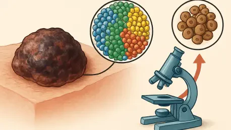
To uncover the mechanisms behind treatment failure, the research team conducted a meticulous analysis of tumor biopsies from the SWOG S1616 clinical trial, specifically focusing on patients whose melanoma had progressed despite receiving initial anti-PD-1 therapy. The methodology involved a multi-modal approach that combined traditional genetic sequencing with high-resolution spatial imaging to create a four-dimensional view of the tumor’s evolution. By taking samples immediately before the start of combination therapy and again approximately one month into the cycle, the researchers could observe real-time changes in the cellular landscape. This allowed them to see not just which genes were “turned on,” but how different types of immune cells moved, interacted, and organized themselves in response to the pharmacological pressure. This high level of detail provided a dynamic look at the cellular battlefield, moving the field of oncology from static snapshots to a comprehensive narrative of immune engagement.
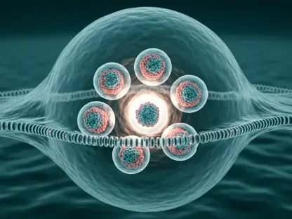
The use of high-resolution imaging technology allowed the scientists to identify “cellular neighborhoods,” which are specific clusters where immune cells and cancer cells interact. This spatial mapping revealed that the density of certain cells is less important than their proximity to one another and to essential structural features like blood vessels. By tracking these interactions across a diverse cohort of patients, the team could correlate specific physical patterns with clinical outcomes, such as tumor shrinkage or continued growth. This approach naturally leads to the conclusion that the physical geography of a tumor is a living, breathing map that dictates the success or failure of modern medicine. These diagnostic tools are now being viewed as the next generation of pathology, offering a way to visualize the effectiveness of a drug long before traditional scans might show a change in tumor size. Such precision allows for a more agile approach to cancer management, where treatment plans can be adjusted based on the micro-environmental shifts observed at the cellular level.
Spatial Architecture and the Future of Treatment
Identifying Success Patterns: The Vital Infrastructure of Immune Recovery
One of the most profound revelations of the study is that the presence of CD8 T cells, often called the “assassins” of the immune system, is only half of the equation for a successful recovery. The researchers found that the most reliable predictor of treatment success is the specific proximity of these T cells to the melanoma cells they are meant to destroy. In patients who responded positively to combination immunotherapy, the T cells were not scattered randomly; instead, they were tightly clustered around the cancer cells in highly organized neighborhoods. These successful clusters were also found to be in close contact with functional blood vessels, which serve as the essential infrastructure for the immune response. These vessels act as highways, allowing a constant stream of fresh immune cells to infiltrate the tumor and providing the metabolic support needed to sustain a prolonged and effective attack against the malignancy.
Furthermore, these responding neighborhoods were characterized by a diverse team of immune cells working in concert, including regulatory T cells and monocytes that provide necessary chemical signals to maintain the offensive. This suggests that a successful immune response is not a solo effort by a single cell type but a sophisticated team maneuver that requires a specific “zoning” of the tumor environment to function. When the tumor is organized in this way, the immunotherapy acts as a catalyst, accelerating a process that the body’s geography is already prepared to support. This spatial insight clarifies why some tumors are “hot” and ready for treatment, while others remain “cold” and inaccessible. Understanding this architecture allows researchers to think about cancer not just as a genetic disease, but as a structural problem that requires the right physical conditions to be solved. This discovery paves the way for new therapies that focus on “re-zoning” the tumor, perhaps by improving blood flow or encouraging T cell clustering to mimic the patterns seen in successful cases.
Personalized Medical Strategies: Remodeling the Tumor Environment for Success
Conversely, the study identified that tumors resistant to treatment often featured a very different spatial configuration, marked by dense and isolated clusters of plasma cells. These clusters appeared to create a barrier or a “cold” zone that effectively excluded T cells from the core of the tumor or rendered them inactive once they arrived. In these non-responding cases, the cancer was able to grow unchecked because the immune system’s primary hunters were physically kept at a distance or lacked the supportive infrastructure of nearby blood vessels and signaling cells. This finding marks a significant paradigm shift toward spatial biology in the field of personalized medicine, suggesting that a biopsy should be used to judge a tumor’s structural readiness for a specific drug. If a biopsy reveals a “disorganized” or “blocked” neighborhood, doctors can immediately recognize that standard immunotherapy is unlikely to work, even if the genetic markers suggest otherwise.
In the coming years, this spatial intelligence will likely lead to the development of “remodeling” strategies, where patients with resistant tumor architectures are given treatments designed to alter the physical layout of the malignancy before or alongside immunotherapy. For instance, targeted therapies or specific types of radiation could be used to break up plasma cell clusters or stimulate the growth of new blood vessels, essentially “priming” the neighborhood for a successful T cell invasion. This approach moves away from the “one-size-fits-all” model of oncology and toward a more nuanced strategy where the treatment is tailored to the unique geography of each patient’s cancer. By focusing on the physical environment, the medical community is moving one step closer to ensuring that advanced skin cancer is no longer a terminal diagnosis for the 70% of patients who currently do not respond to standard combination therapies. The goal is to transform the tumor from a resistant fortress into an accessible target that the immune system can finally dismantle.
The UCLA study demonstrated that the spatial organization of immune cells serves as a more accurate predictor of immunotherapy success than genetic analysis alone. Clinical teams now recognize that the proximity of CD8 T cells to cancer cells and the presence of supportive vascular infrastructure are the primary hallmarks of a responsive tumor. These findings established a clear link between the physical “neighborhood” of a malignancy and the ultimate clinical outcome for the patient. To improve survival rates in advanced melanoma, future research must focus on developing pharmaceutical and radiological interventions that can actively remodel the tumor microenvironment. By transforming the structural layout of resistant tumors to mimic successful spatial patterns, oncologists can potentially expand the reach of life-saving immunotherapies to a much larger population. The path forward involves integrating high-resolution spatial mapping into routine diagnostic protocols to ensure that every patient receives a treatment strategy aligned with the physical reality of their disease.