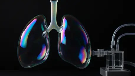

Respiratory motion is not just a byproduct of breathing but a fundamental driver of how the human immune system responds to viral threats. For decades, the medical community struggled to understand why laboratory experiments involving lung infections often failed to match the complex reality of a living patient. This gap exists because the lung is not merely a static filter for gas exchange; it is a dynamic organ that experiences continuous mechanical strain as the chest cavity expands and contracts. Replicating this constant motion within a controlled environment has remained one of the most significant hurdles in bioengineering. Traditional methods, such as growing cells in a petri dish, completely strip away the physical context that dictates how cells function and defend themselves. As researchers look for better ways to predict disease outcomes and test new treatments, the need for a system that truly mimics the rhythmic life of a human lung has become increasingly urgent for global health security and the development of future-ready medicines.
Limitations of Previous Laboratory Models
Before the breakthrough at the Pohang University of Science and Technology, researchers were forced to rely on rudimentary “lung-on-a-chip” devices that frequently missed the mark. These chips typically utilized synthetic materials like polydimethylsiloxane, which offered transparency and flexibility but lacked the essential biochemical cues that human cells require to thrive. Because these materials were inherently non-biological, they often skewed the results of pharmacological tests, leading to inaccurate predictions of how a drug might behave in a real patient. Furthermore, these static models failed to incorporate the three-dimensional architecture of the alveoli, which are the millions of tiny air sacs responsible for life-sustaining oxygenation. The lack of physiological relevance meant that scientists could only observe how cells behaved in a vacuum, ignoring the critical interplay between mechanical forces and cellular biology that defines the respiratory system daily.
Attempts to move toward more realistic biological scaffolds using hydrogels initially met with significant frustration in various laboratory settings. While hydrogels are much softer and better suited for cell growth than rigid plastic or silicone, they were traditionally too delicate to withstand the repetitive stretching that occurs during inhalation and exhalation. When researchers tried to simulate breathing cycles, these fragile membranes would often rupture or degrade long before meaningful data could be collected. This durability issue prevented long-term studies of chronic respiratory conditions or slow-acting viral infections that require days or weeks of observation. Consequently, the scientific community remained stuck between choosing a durable but inaccurate synthetic material or a realistic but fragile biological one. Breaking this deadlock required a total rethink of how thin membranes are constructed, leading engineers to look toward the physics of common soap bubbles for a brand new answer.
Engineering a Resilient Hydrogel Membrane
The research team found a source of inspiration in the physical mechanics of soap bubbles, which maintain a delicate balance between surface tension and structural elasticity. By observing how soap films can stretch and deform without breaking, they developed a technique to create a composite hydrogel membrane that is both incredibly thin and remarkably resilient. Using a specialized dipping process, a mold was submerged into a precisely balanced solution of polymers and monomers and then slowly withdrawn. This method allowed for the formation of a uniform, high-strength film that mimics the physical properties of human alveolar tissue. Unlike previous attempts, this new membrane could be engineered to a thickness comparable to the biological walls found in the lung, providing a realistic barrier for gas exchange while maintaining enough structural integrity to function under pressure. This innovation represented a major leap forward in both material science and modern bioengineering.
To ensure the longevity and reliability of this new platform, the engineers subjected the soap-bubble-inspired membrane to rigorous mechanical stress tests that would have destroyed earlier models. They designed an actuation system that replicates the negative pressure environment created by the human diaphragm, pulling on the hydrogel to simulate the intake of air. The results were unprecedented, as the artificial lung successfully completed approximately 240,000 breathing cycles without any signs of failure or material fatigue. This level of endurance allows for the study of disease progression over extended periods, providing a stable environment for cells to grow and respond to external stimuli. By matching the mechanical resilience of native lung tissue, the device offers a reliable window into the long-term effects of environmental pollutants or the gradual recovery from an infection. This technological foundation set the stage for adding the complex biological layers necessary for a truly functional model.
Biological Integration and Future Therapeutic Applications
Creating a structural membrane was only the first step; the researchers then had to populate it with living human cells to mirror the intricate architecture of the alveoli. Utilizing advanced 3D bioprinting technology, the team meticulously layered different types of cells to recreate the alveolar-capillary barrier. This barrier is essential for health, as it separates the air we breathe from the blood circulating through our bodies. The bioprinting process allowed for the precise placement of vascular cells on one side of the hydrogel, followed by a basement membrane and a layer of epithelial cells on the other. This three-layered approach ensures that the cells can communicate with one another just as they do in the human body, allowing for a more accurate representation of how nutrients and signals pass through the lung wall. By controlling the spatial distribution of these cells, the researchers created a high-fidelity biological interface that responds dynamically to the environment.
Looking ahead, the transition from conventional testing to these dynamic, soap-bubble-inspired platforms offered a clear path toward more ethical and effective medical research. In 2026, the scientific community began to prioritize these high-fidelity models to reduce the historical reliance on animal testing, which often fails to replicate human-specific immune responses. The ability to use patient-derived cells within these breathing systems allowed for the development of personalized treatment plans, where a drug’s efficacy could be verified on a specific individual’s tissue before it was ever administered. Researchers were encouraged to integrate these systems into standard laboratory workflows to capture the mechanical variables that were previously ignored. This shift towards biomimetic technology ensured that the next generation of therapeutics was grounded in the true physical reality of the human body. The integration of mechanical force and biological layering ultimately transformed how scientists approached the complexities of respiratory medicine.