
The internal chronology of the human brain appears to be a negotiation between hardwired genetic programs and the external environment rather than a purely reactive state. This realization marks a significant departure from traditional neurobiology, which often viewed the brain as a plastic organ almost entirely shaped by the sensory world. For years, scientists struggled to replicate the slow, majestic pace of human neural development in the laboratory, as most cultures withered long before they reached anything resembling true maturity. However, a groundbreaking study has recently upended these limitations, proving that with the right bioengineering support, human brain tissue can thrive and evolve for nearly seven years in a controlled setting. These peppercorn-sized clusters, known as organoids, have demonstrated an incredible degree of biological autonomy, ticking away through the years without a heart to pump blood or a nervous system to provide feedback. This longevity allows researchers to observe the molecular shifts that occur as a brain moves from its embryonic origins toward the complexity of early childhood, revealing that our developmental tempo is etched deeply into our DNA.
The Architecture and Evolution of Synthetic Brain Tissue
Mapping the Growth: The Cerebral Cortex Model
The primary focus of this intensive research centered on the cerebral cortex, which serves as the seat of higher human cognition and complex behavior. Unlike traditional two-dimensional cell cultures that offer only a flat, distorted view of cellular interaction, these three-dimensional organoids were engineered to mimic the actual architecture of the human neocortex. By starting with human pluripotent stem cells and applying a sophisticated series of biochemical signals, the team successfully directed the cells to adopt a dorsal cerebral cortex identity. This process was not merely about simple growth but about complex organization; the cells moved, layered, and connected in a manner that mirrored the natural biological scaffolds found in a developing fetus. The resulting tissue provides a high-fidelity model that researchers can manipulate and observe in real-time, offering a level of access that is impossible with living human subjects. This transition to long-term 3D modeling represents a significant leap forward in our ability to simulate the most complex organ in the known universe.
Each organoid produced in this study contained upwards of one million cells, forming a dense and intricate network of neural progenitors and maturing neurons. The most striking observation was that these clusters followed a predictable, human-specific chronological sequence of development without any external cues. In the natural world, a child’s brain is bombarded with hormones, nutrition, and sensory data, all of which were absent in the sterile environment of the laboratory dish. Despite this isolation, the organoids did not stall or grow erratically; instead, they matured with a rhythm that was unmistakably human. This suggests that the early stages of brain construction are guided by a robust, internal genetic program that operates on a much slower timeline than that of other mammals. By documenting this process over several years, the researchers have effectively created a biological time-lapse, allowing us to see how the architecture of the mind is built, piece by piece, according to an ancient and internal blueprint that resides within each individual cell.
Longitudinal Analysis: Molecular Benchmarking Over Time
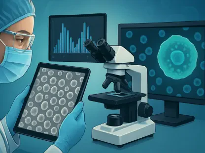
To quantify the progress of these synthetic tissues, the research team undertook a massive longitudinal analysis that involved tracking more than 400,000 individual cells over a multi-year period. Utilizing single-cell RNA sequencing, they were able to map gene activity across sixteen distinct timepoints, starting from the first fifteen days of growth and extending through five years of maturation. This data provided a granular view of how different cell types emerge and how their genetic expressions shift as the tissue ages. By comparing these lab-grown sequences to established reference sets of actual human brain tissue, the researchers confirmed that the organoids were not merely surviving but were actively following the human developmental timeline with startling precision. This molecular benchmarking is crucial, as it validates the organoid as a legitimate proxy for human biology, ensuring that the insights gained in the lab are applicable to the understanding of real human health and the progression of neurodevelopmental milestones.
The comparative data revealed that organoids aged under two months almost perfectly mirrored the gene expression patterns of first-trimester human fetal tissue. As the organoids reached the one-year mark, they began to express gene programs that are typically associated with the brains of postnatal children. This alignment persisted throughout the study, demonstrating that the cells possess an inherent knowledge of their age and the specific genes they should be activating at any given moment. Such findings are revolutionary because they suggest that the protracted maturation process of the human brain is a cellular property rather than a systemic one. By establishing this high-fidelity molecular timeline, the researchers have provided a roadmap for future studies, allowing scientists to pinpoint exactly where developmental trajectories might diverge in cases of disease or genetic mutation. This offers a path toward studying human-specific biology within a laboratory setting that requires no invasive procedures or unethical experimentation on living subjects.
Biological Time and Cellular Memory
Deciphering the Clock: The Internal Epigenetic Mechanism
Beyond the observation of gene activity, the study delved into the underlying mechanisms of biological aging by utilizing advanced epigenetic clocks. These clocks measure the biological age of tissue by analyzing DNA methylation, which consists of specific chemical marks on the genome that predictably change as an organism matures. By examining these genomic regions, the researchers found that they could determine the age of an organoid with remarkable accuracy, matching the ticking of the lab-grown tissue to the chronological time it had spent in the incubator. This discovery indicates that human cells possess an internal odometer that continues to record the passage of time even when removed from the context of a whole organism. It suggests that the aging process is a fundamental, coordinated chemical program that is largely independent of the external environment, providing a new perspective on how our bodies track their own lifespan at the molecular level without systemic cues.
The existence of this internal clock within isolated brain tissue has profound implications for our understanding of neurobiology and the nature of aging. It implies that the pace of our cognitive maturation is not something that purely happens to us, but something that is driven from within our cells. While the circulatory and nervous systems certainly play roles in supporting and refining the brain, the primary rhythm of development is maintained by a series of chemical shifts within the DNA itself. This intrinsic timing mechanism explains why human development is so much slower and more deliberate than that of other species; our cells are programmed to take their time to ensure complexity. By mastering the ability to read these epigenetic clocks in a lab-grown model, scientists can now begin to investigate what happens when the clock runs too fast or too slow, potentially identifying the molecular triggers for premature aging or developmental delays that were previously invisible to researchers.
Temporal Memory: The Resilience of Cellular Identity
One of the most fascinating aspects of the study involved an experiment designed to test whether neural progenitor cells could remember their developmental age. Researchers created chimeric organoids by mixing old neural progenitors, which had already aged for nine to twelve months, with young progenitors that were only fifteen days old. The goal was to see if the environment provided by the younger cells or the culture medium could reset the older cells, forcing them to repeat early developmental stages. However, the results demonstrated a powerful phenomenon known as temporal memory. The older cells did not revert to an earlier state; instead, they maintained their original trajectory, skipping the early steps they had already completed and continuing to produce late-stage neurons. This suggests that once a cell reaches a certain point in its developmental history, its identity and progress are remarkably durable and resistant to external environmental changes or signals.
This finding of temporal memory underscores the autonomy of human biological programs and suggests that the history of a cell is just as important as its current environment. While the younger cells in the mixture were able to influence the older ones to remain active and proliferative for longer periods, they could not overwrite the molecular knowledge the older cells had already acquired. This molecular state appears to be dictated by the cell’s own internal record of its past, ensuring that development proceeds in a logical, one-way direction. For medical research, this is a double-edged sword; it means that while cells are resilient, they may also be stubborn, making it difficult to reset tissues that have already begun a path toward disease. However, it also provides a unique opportunity to study how cellular identity is maintained over years, offering clues into how we might one day protect healthy brain cells from the degrading effects of time and external stressors.
Sustaining Vitality: Neuronal Health and Electrical Activity
Maintaining fragile neurons over several years is a significant technical challenge in biotechnology that requires precise environmental control. To ensure the survival of these cells, the team used a specialized activity-permissive medium that encouraged the development of elaborate synaptic structures. This environment allowed the organoids to exhibit spontaneous electrical activity for at least two years, a sign that the neurons were not only alive but were also forming functional connections. These electrical pulses are vital signs of healthy, maturing tissue, yet the researchers were careful to emphasize that this activity is a fundamental biological property. It does not equate to consciousness or thought, as the organoids lack the necessary sensory input and a complex nervous system to process information in a meaningful way. Instead, the activity serves as a marker of cellular health and the successful integration of neural networks.
The ability to sustain electrical activity over such a long duration provides a stable platform for studying how neural communication changes as the brain ages. In traditional models, researchers often see a decline in cellular health before they can observe long-term synaptic changes. By providing an environment where these processes can play out over years, the study has opened a window into the endurance of human neurons. This allows for the investigation of how synaptic pruning and network refinement occur in a human context, free from the noise of a complete biological system. Understanding the factors that keep these neurons electrically active and healthy for years in a dish could lead to new strategies for preserving brain function in aging populations. It also highlights the incredible robustness of human neurons, which can maintain their functional integrity for half a decade with only minimal biochemical support from their surrounding medium.
Future Horizons in Medical Research
Modeling Progress: Neurodevelopmental and Degenerative Disorders
The human cerebral cortex requires nearly twenty years to reach full maturity, a timeline that far exceeds that of any laboratory animal typically used in medical research. This discrepancy has long made it difficult to study human-specific conditions that emerge during late-stage development or early adulthood. By extending the reproducible culture window for brain organoids to seven years, scientists have effectively bridged a massive gap in our research capabilities. We can now study the origins of complex disorders such as epilepsy, autism, and certain forms of schizophrenia in a human-specific context over a meaningful span of time. These conditions often involve subtle molecular shifts that do not manifest until years after birth, making them nearly impossible to capture in short-term studies. Having a stable, multi-year human model allows researchers to observe how these diseases unfold across the developmental timeline, potentially leading to earlier interventions.
Furthermore, this long-term modeling capability provides a new lens through which to view neurodegenerative diseases and the general process of brain aging. Since the organoids follow a human-specific aging schedule, researchers can introduce genetic variances associated with conditions like Alzheimer’s or Parkinson’s and observe how these alterations change the brain’s trajectory over several years. This offers a way to test how specific genes interact with the passage of time, providing insights into why some individuals are more resilient to aging than others. The ability to watch a human brain model age in a dish for half a decade allows for the kind of controlled, longitudinal observation that was previously a distant dream. This approach naturally leads to a more personalized form of medicine, where a patient’s own cells could be used to grow organoids that predict their future neurological health, allowing for proactive rather than reactive medical care starting from the earliest stages of life.
The Road Ahead: Accelerating Discovery and Therapeutic Testing
While the longevity of these organoids represented a historic scientific milestone, the research team focused on refining these tools for more efficient use in the pharmaceutical industry. After the temporal memory of progenitor cells was better understood, scientists explored methods to fast-forward organoids to later developmental stages without the need to wait several years for them to grow naturally. By searching for the chemical keys to advance the epigenetic clock, researchers aimed to create mature brain tissue models in a fraction of the time, enabling high-throughput testing of drugs and genetic therapies. This approach promised to drastically accelerate the pace of discovery, allowing for the rapid screening of compounds that could protect the brain or correct developmental errors. The project transformed these organoids from experimental curiosities into high-fidelity diagnostic tools that were eventually deployed across the global medical research landscape.
The study ultimately concluded that the fundamental sequence of human brain development was a journey that cells were inherently prepared to take on their own terms. The researchers demonstrated that while the external environment remained necessary for survival and refinement, the core tempo of our species’ cognitive growth stayed a hardwired property of human biology. This effort provided a profound new perspective on the resilience and autonomy of human cells, highlighting their ability to maintain a complex developmental schedule for over half a decade in a plastic dish. As progress continued from 2026 into a new era of biotechnology, these insights served as the foundation for a deeper understanding of what made the human brain unique. By bridging the gap between isolated cellular biology and the mystery of the aging process, this research paved the way for a future where the secrets of our longest-lived and most complex organ were finally within reach to understand and protect.