
Identifying CD31 protein expression in engineered tissue constructs serves as a vital indicator that a functional internal blood vessel network has begun to take shape. For patients grappling with catastrophic bone injuries or the aftermath of tumor removals, the quest for a permanent solution often ends in frustration due to the limitations of current grafting methods. While the body possesses an innate ability to mend minor fractures, massive structural voids remain a persistent challenge that standard medicine struggles to bridge. Recent advancements have shifted the focus toward a more sophisticated biological paradigm, where researchers at Penn State are now leveraging the power of “genetic switches” to orchestrate complex repairs. By programming stem cell clusters to assume specific roles, science is moving closer to creating living bone that behaves exactly like its natural counterpart. This method represents a departure from static scaffolds, offering a dynamic way to heal bone that requires more than just a mineral filler to survive.
Overcoming the Biological Limits of Engineered Tissue
The Challenge: Navigating the Diffusion Limit
The primary obstacle in the engineering of substantial bone grafts remains the physiological constraint known as the “diffusion limit.” In many traditional tissue engineering models, cells located more than 200 micrometers from a nutrient source quickly become hypoxic and die, leading to the eventual necrotic failure of the entire implant. This fundamental bottleneck has long prevented the development of thick, clinically relevant tissue structures, as the core of the graft remains isolated from the host’s circulatory system. Instead of relying on the slow, passive migration of host blood vessels into the graft, modern strategies focus on pre-vascularizing the construct. By building the infrastructure for blood delivery into the tissue during the manufacturing process, researchers can ensure that oxygen and essential nutrients reach the innermost layers. This proactive management of the cellular environment prevents central cell death and sets the stage for a more robust integration with the body.
Addressing this systemic failure requires a “heterocellular” strategy that moves beyond the simplistic view of bone as a rigid mineral mass. By integrating multiple cell types into a single engineered construct, scientists can foster a microenvironment that supports both skeletal structure and vascular health simultaneously. This integrated approach ensures that as the new bone matrix begins to mineralize, a corresponding network of capillaries is already forming to sustain it. The success of this method depends on the ability to program these cells to work in harmony, preventing the common issue where one tissue type outcompetes another for resources. Consequently, the engineered graft acts more like a self-contained biological ecosystem rather than a foreign object. This shift in perspective is essential for the creation of large-scale repairs that can withstand the metabolic demands of human physiology. Establishing such a functional internal blood supply is the difference between a temporary patch and a lifelong medical solution.
MicroRNA Signaling: Programming Cellular Fate
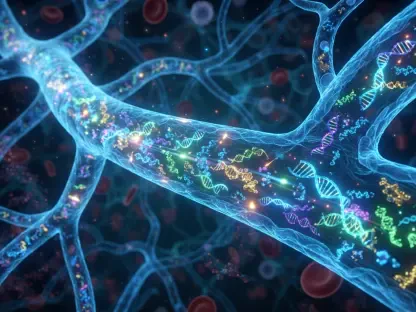
The engine driving this biological coordination is the strategic use of microRNA molecules, which serve as highly specific genetic switches within the cell. These small, non-coding RNA strands do not alter the genetic code itself but rather dictate which proteins the cell produces, effectively steering stem cells toward specific developmental paths. For instance, the use of miR-148b is employed to trigger the differentiation of stem cells into osteoblasts, which are the primary cells responsible for bone formation. Simultaneously, miR-210 is utilized to stimulate the expression of factors that promote angiogenesis, the growth of new blood vessels. By delivering these molecular instructions precisely, researchers can transform a uniform population of undifferentiated cells into a specialized workforce capable of complex tissue assembly. This level of control allows for the creation of a sophisticated “blueprint” where every cell knows its role before it is ever placed into a clinical setting.
Rather than utilizing individual cells, which can easily lose their signaling potency or drift away from the target site, the team organizes these programmed cells into three-dimensional clusters known as spheroids. These spheroids function as miniature tissue building blocks, providing a much richer environment for cell-to-cell communication and structural support. The dense cellular arrangement within a spheroid mimics the natural density of human tissue, allowing for more robust signaling and better survival rates during the bioprinting process. Furthermore, the use of spheroids enables the precise mixing of different cell types within a single construct, ensuring that bone-promoting and vessel-promoting units are always in close proximity. This spatial arrangement is critical because the growth of bone and blood vessels is naturally interdependent; one cannot thrive without the other. By starting with these biologically primed clusters, the resulting engineered tissue is far more resilient and capable of rapid maturation.
Advanced Manufacturing and Structural Design
Precision Bioprinting: Organizing Cellular Units
To transform these specialized spheroids into a cohesive and functional bone graft, researchers utilize a sophisticated technique known as aspiration-assisted bioprinting. Unlike traditional extrusion-based bioprinting, which often subjects cells to high levels of shear stress, this method employs controlled vacuum pressure to “pick and place” individual spheroids with remarkable accuracy. This precision allows for the construction of highly organized tissue architectures that would be impossible to achieve through manual methods or less refined printing technologies. By placing each bone-promoting and vessel-promoting spheroid at specific coordinates, the system can create a complex, interleaved pattern that optimizes biological signaling across the entire construct. This level of spatial control is vital for ensuring that no part of the graft is left without the necessary instructions for growth. The ability to manipulate individual cellular units at this scale represents a significant leap forward in the field of regenerative medicine.
The structural integrity of the bioprinted graft is maintained by a specialized microgel scaffold that holds the spheroids in place as they begin to fuse. This scaffold provides the immediate physical support required to bridge large bone defects, but it is designed to be temporary, eventually giving way to the natural matrix produced by the cells themselves. Within this controlled environment, the strategically placed spheroids begin to communicate across the gaps, extending their cellular reach to form a single, integrated unit. This process of fusion is critical for the long-term success of the repair, as it ensures that the mechanical load is distributed evenly across the new bone structure. By carefully managing the distance between different cell clusters, the bioprinting process ensures that the signaling molecules reach their targets efficiently. This meticulous organization prevents the formation of weak spots or non-functional regions, resulting in an engineered graft that is both biologically active and mechanically sound from the moment of implantation.
Integrated Architecture: Creating a Living System
The overarching goal of this precise arrangement is to mimic the complex, multi-layered architecture found in natural human bone. Human skeletal tissue is not a uniform block; it is a highly specialized system of mineralized matrix, nerves, and intricate vascular networks. By bioprinting specialized spheroids into a heterogeneous pattern, researchers are able to recreate this complexity on a macroscopic scale. This engineered environment allows the bone-forming cells to receive constant metabolic support from the emerging blood vessel network, which in turn accelerates the mineralization process. Furthermore, the interaction between these different cell types creates a feedback loop that enhances the overall vitality of the graft. As the vessels mature, they release growth factors that further stabilize the developing bone tissue, creating a self-sustaining cycle of regeneration. This level of integration is what allows the engineered construct to transition from a laboratory-grown model to a functional piece of living anatomy.
Beyond the immediate biological benefits, this integrated design addresses the critical need for long-term stability in large-scale bone repairs. When a graft is successfully vascularized, it gains the ability to remodel itself in response to physical stress, a hallmark of healthy bone. Without this internal blood supply, even the strongest synthetic scaffold will eventually become brittle and fail because it cannot repair the microscopic damage that occurs during daily activity. The bioprinted constructs are designed to be dynamic, allowing for the natural turnover of cells and minerals that keeps bone healthy over time. By establishing these biological systems from the outset, the research team is providing a pathway for permanent recovery that standard bone grafts simply cannot match. This approach treats bone repair not as a mechanical assembly task, but as the cultivation of a complex living system. Consequently, the final structure is better equipped to integrate with the host’s existing skeleton and provide lasting support for the patient’s physical needs.
Validating the Success of Genetic Programming
Laboratory Evidence: Synergistic Tissue Maturation
The effectiveness of using genetic switches to drive tissue development was first validated through rigorous laboratory testing, which tracked the expression of specific genetic markers. Researchers observed that when stem cells were treated with the designated microRNAs, they consistently produced the proteins necessary for their specialized roles. For example, the cells programmed for bone growth showed high levels of alkaline phosphatase and osteocalcin, which are essential for creating the hard mineral matrix of bone. Meanwhile, the vessel-promoting cells exhibited robust expression of vascular endothelial growth factor, signaling their readiness to form capillary structures. Interestingly, the data revealed that these two cell populations performed better when grown together than they did in isolation. This synergy suggests that the biological signals sent between the bone and vessel units create a more favorable environment for maturation. This mutual reinforcement is a key discovery, confirming that the dual-track programming strategy is biologically sound and efficient.
Furthermore, the physical maturation of the bioprinted constructs was monitored to ensure that the spheroids were successfully fusing into a single, cohesive tissue. Over the course of the incubation period, the individual clusters of cells merged, forming a continuous network of mineralized tissue interspersed with developing vascular channels. Microscopic analysis confirmed that the “genetic switches” remained active throughout this process, guiding the cells as they matured from undifferentiated precursors into specialized functional units. The resulting tissue displayed a level of organization and density that closely mirrored natural trabecular bone. This laboratory success provided the necessary proof of concept to move forward with animal trials, as it demonstrated that the engineered constructs could maintain their identity and function within a complex three-dimensional environment. The ability to observe these specific biological markers in vitro offered a clear roadmap for what to expect during the clinical integration phase, marking a significant milestone in the development of the project.
Animal Model Results: Achieving High Recovery Rates
The most compelling evidence for the potential of this technology came from studies conducted on animal models with critical-sized bone defects. In these experiments, mice with significant skeletal injuries were treated with the bioprinted, genetically programmed grafts to evaluate their healing capacity. Within just six weeks, the animals that received the dual-spheroid constructs showed a remarkable 93 percent recovery rate, far exceeding the performance of control groups that received standard scaffolds or single-cell type grafts. This high success rate was attributed to the presence of the pre-programmed vascular network, which allowed the new bone to survive and integrate almost immediately upon implantation. Imaging studies showed that the engineered tissue had not only filled the void but had also begun to fuse with the host’s original bone. This rapid integration is essential for restoring full mobility and preventing the displacement of the graft. The results underscored the importance of simultaneous bone and vessel growth.
Crucially, the analysis of the recovered tissue revealed high levels of CD31, a protein that serves as a definitive marker for functional blood vessels. The presence of this protein confirmed that the internal vascular network bioprinted into the graft had successfully connected with the host’s circulatory system. This connection allowed the graft to receive a steady supply of blood, ensuring its long-term viability and allowing it to maintain its mineral density over time. In contrast, the control groups showed significant signs of bone resorption and cell death in the center of the grafts, illustrating the fatal consequences of poor vascularization. The ability of the genetically programmed cells to establish a living, breathing blood supply within the bone matrix is what sets this research apart from previous attempts at large-scale tissue engineering. By proving that these “genetic switches” can translate into real-world healing, the study has paved the way for a new generation of regenerative therapies that address the body’s most complex repair challenges.
Clinical Outlook and Research Trajectory
Targeting TraumBeyond Standard Bone Healing
The current focus of this research is specifically on “critical-sized defects,” which are injuries so large that the body’s natural regenerative mechanisms are unable to heal them. These types of injuries often arise from high-impact trauma, the surgical removal of large bone tumors, or the destruction of tissue due to chronic and aggressive infections. In such cases, the patient is typically left with a permanent void that requires a massive graft, which often carries a high risk of failure or rejection. By providing a pre-vascularized, genetically optimized graft, this technology offers a way to restore structural integrity to even the most severely damaged limbs. The goal is to provide surgeons with a reliable, off-the-shelf solution for cases where traditional autografts—taking bone from another part of the patient’s body—are not feasible or would cause too much additional trauma. This shift toward “living” implants could drastically reduce the need for multiple follow-up surgeries for trauma victims.
As the technology matures, it is expected to provide a new standard of care for complex orthopedic and reconstructive procedures. While standard fractures can usually be managed with casts or simple internal fixation, massive bone loss requires a different biological approach that addresses the lack of a local blood supply. The ability to engineer tissue that brings its own vascular system to the injury site is a game-changer for reconstructive surgery. Beyond just filling a hole, these grafts are designed to become a permanent part of the patient’s skeletal system, capable of bearing weight and adapting to the physical demands of life. This represents a fundamental move away from viewing bone as a simple mechanical support toward understanding it as a complex organ that requires integrated biological management. By focusing on these high-stakes clinical scenarios, the research team is addressing some of the most difficult challenges in modern orthopedics, offering hope for patients who previously had very few options.
Future Directions: Safety and Scaling the Technology
Looking forward, the next critical steps involve transitioning the research from small animal models to larger animals that more closely mimic human physiology and mechanical loading. These larger-scale trials will be essential for verifying that the bioprinted constructs can maintain their structural integrity and vascular function under the significant stresses placed on human limbs. Researchers also need to establish rigorous safety protocols regarding the long-term use of microRNA-modified cells. While the genetic switches are highly effective at guiding development, it is vital to ensure that the programmed cells remain stable and do not exhibit any unintended behaviors over several years. This will involve detailed monitoring of cell lineage and genetic stability within the host environment. Additionally, the manufacturing process must be refined to ensure that bioprinted grafts can be produced consistently and cost-effectively for clinical use. Standardizing the production of specialized spheroids will be a major focus.
The ultimate success of this approach will depend on the ability to customize these treatments for individual patients, potentially using their own stem cells to avoid any risk of immune rejection. This personalized medicine model would involve harvesting a patient’s cells, programming them with the necessary genetic switches, and then bioprinting a custom-shaped graft to fit their specific injury. While this adds a layer of complexity to the process, it offers the best possible chance for seamless integration and long-term health. The research team is also exploring ways to incorporate other tissue types, such as nerves and cartilage, into the bioprinted structures to create even more comprehensive repair solutions. This holistic approach to tissue engineering could eventually lead to the replacement of entire segments of bone and joint systems with living, functional biological units. As the industry moves closer to these milestones, the emphasis will remain on ensuring that these advanced technologies are both safe and accessible to those who need them most.
The successful integration of genetic switches and precision bioprinting established a new benchmark for the field of regenerative medicine. By moving away from inert scaffolds and toward dynamic, pre-vascularized tissue, scientists demonstrated that large-scale bone repair was a biological challenge that could be solved through engineering. This research highlighted the critical importance of vascularization in the survival of thick tissue grafts, proving that internal blood supplies were the key to long-term structural success. The high recovery rates observed in initial trials provided the necessary confidence to push the technology toward more complex clinical applications. In the following years, the focus shifted toward large-scale manufacturing and the creation of personalized grafts tailored to specific patient anatomies. This progression required a collaborative effort between biologists, engineers, and surgeons to translate laboratory findings into a practical medical reality. Ultimately, the development of these “living” bone grafts offered a transformative solution for those suffering from severe skeletal trauma and paved the way for future innovations in whole-organ biofabrication.