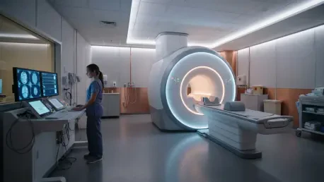

The shift toward non-invasive kinetic quantification allows clinicians to measure tracer behavior without the logistical burdens and patient discomfort associated with manual blood sampling. In the current medical landscape of 2026, the widespread adoption of Long Axial Field of View (LAFOV) scanners has fundamentally transformed the practice of nuclear medicine. These sophisticated systems provide a comprehensive and simultaneous view of the entire human body, allowing for the real-time capture of dynamic physiological processes across all major organ systems. This technological leap addresses the long-standing limitations of traditional Positron Emission Tomography, which historically required multiple bed positions and significantly longer scan times to cover comparable anatomical areas. By capturing the total body in a single acquisition window, clinicians can now observe systemic interactions and metabolic pathways that were previously obscured. The increased sensitivity of these scanners also allows for substantial reductions in radiopharmaceutical dosages, enhancing safety for pediatric and chronic care patients.
Advancing Non-Invasive Quantitative Imaging and Vascular Diagnostics
The emergence of total-body imaging has catalyzed a shift toward sophisticated quantitative analysis that was once reserved for high-end research environments. By providing a continuous view of the circulatory system, these scanners enable the precise tracking of radiotracers as they move through the heart and major arteries. This capability is the foundation for non-invasive kinetic modeling, which provides a deeper understanding of disease behavior and treatment response. As these systems become more integrated into routine hospital workflows, the focus has shifted toward refining the software algorithms that translate this raw data into actionable clinical insights. This transition ensures that every patient can benefit from the high-precision diagnostics that were previously limited by the technical complexity of manual blood sampling and arterial monitoring. This evolution is particularly visible in the management of complex oncological cases and the burgeoning field of vascular molecular imaging.
Transforming Major Vessels: Virtual Blood Sampling
The validation of image-derived input functions (IDIF) represents a milestone in the effort to make quantitative PET imaging more accessible and patient-friendly. Recent clinical data from 2026 confirms that the descending aorta can effectively serve as a “virtual” blood sample, providing the necessary data for kinetic modeling without the need for invasive arterial lines. This method leverages the high spatial resolution of total-body scanners to extract precise tracer activity directly from the vessel, which eliminates the logistical bottlenecks and physical discomfort associated with manual sampling.
By removing the requirement for an indwelling arterial catheter, medical centers have streamlined their diagnostic pipelines and improved overall patient throughput. This advancement is especially impactful in oncology, where the ability to generate metabolic maps non-invasively allows for more frequent and detailed monitoring of tumor progression. The move toward virtual sampling ensures that the benefits of quantitative imaging are available to a broader range of patients, including those for whom traditional invasive procedures might pose a significant risk.
Exploring the Limits: Vascular and Neuroimaging
In the realm of vascular medicine, researchers are successfully using oxygen-15 labeled water to quantify muscle perfusion in patients suffering from peripheral arterial diseases. This technique allows for the non-invasive measurement of blood flow through the extremities, providing essential data for evaluating the effectiveness of revascularization and other surgical interventions. However, the high precision required for imaging smaller vessels in the limbs necessitates advanced motion-correction algorithms, as even minor patient movement can impact the quantitative accuracy of the perfusion maps.
Neuroimaging also continues to benefit from these advancements, as researchers investigate the potential for venous blood samples to substitute for arterial data when using metabolically active tracers. While some findings for specific markers have highlighted the complexity of tracer metabolism in the brain, these efforts are setting vital benchmarks for future technological refinements. The ongoing development of high-fidelity tracers and improved software ensures that the medical community is moving closer to a fully non-invasive neurological assessment that captures the subtle interactions between the brain and the rest of the body.
Standardizing Performance and Integrating Digital Innovation
For total-body PET to remain a cornerstone of modern diagnostics, the data generated across different hospitals and scanner models must be strictly standardized. This ensures that a patient’s results are comparable over time and across different geographic regions, which is essential for global clinical trials and universal treatment guidelines. Current state-of-the-art systems have demonstrated exceptional performance, yet the industry is actively pushing for updated accreditation standards that reflect the superior sensitivity of LAFOV technology. This push for standardization is occurring alongside a rapid digital transformation, where artificial intelligence and machine learning are being used to manage the massive datasets produced by total-body acquisitions. The synergy between high-performance hardware and intelligent software is creating a more efficient and accurate diagnostic environment, allowing clinicians to focus their expertise on the most complex patient needs while delegating routine data processing to automated systems.
Establishing New Protocols: Global Consistency
Ensuring global consistency in PET imaging is a primary goal for the nuclear medicine community as they move toward the adoption of EARL3 standards. These updated protocols are specifically designed to leverage the unique capabilities of total-body systems, such as their extreme sensitivity and wide coverage. By establishing new benchmarks for image quality and quantification, the industry ensures that the data remains reliable and reproducible across various healthcare systems. This foundation of trust is necessary for the development of multi-center research projects and the implementation of standardized patient care pathways.
The move toward EARL3 involves not only software updates but also the creation of new physical calibration tools that can accurately represent the total volume of the human body. This comprehensive approach to quality assurance guarantees that the high-level performance of LAFOV scanners is translated into consistent diagnostic outcomes for every patient. As these standards become the norm throughout 2026 and 2027, the medical community will be better equipped to share data and collaborate on the next generation of life-saving treatments for cancer and neurological disorders.
Streamlining Diagnostics: Artificial Intelligence
The digital frontier of nuclear medicine is currently being reshaped by the integration of Large Language Models and automated reporting tools. Pilot studies have shown that AI can generate structured and accurate clinical reports for melanoma and other cancers, often matching the quality of drafts produced by senior radiologists. By automating these descriptive tasks, medical professionals can significantly reduce their administrative burden, allowing them to dedicate more time to interpreting complex findings and discussing treatment options with their patients.
In addition to reporting, AI is serving as a powerful decision-support tool that helps clinicians identify subtle lesions that might be overlooked in the large datasets generated by total-body scans. These algorithms are trained to recognize patterns of disease across the entire body, providing a second layer of scrutiny that enhances diagnostic sensitivity. This partnership between human expertise and machine intelligence is essential for managing the increased complexity of total-body data, ensuring that every scan provides the most complete and accurate information possible for the patient’s care team.
Exploring Positronium Lifetime: A New Contrast Tool
A groundbreaking development in imaging physics is the emergence of positronium lifetime imaging, which offers a new way to visualize the molecular environment of human tissues. Unlike traditional PET that measures tracer concentration, this method measures the decay time of positron-electron pairs, which is influenced by local chemical conditions such as oxygen concentration. This breakthrough allows for the visualization of the tumor microenvironment in a way that was previously impossible, providing insights into the specific chemical characteristics of cancerous versus healthy tissue.
The high sensitivity of total-body PET scanners is a prerequisite for this technique, as it requires the collection of a large number of decay events to calculate accurate lifetimes. As the temporal resolution of detectors continues to improve throughout 2026, the transition of positronium imaging from the laboratory to the clinic is becoming more likely. This would lead to the development of multiparametric scans, where metabolic and chemical data are combined into a single diagnostic image. This new dimension of biological information will be crucial for the next generation of personalized and targeted therapies.
The Future: Patient-Centered Molecular Imaging
The shift toward total-body systems has fostered a more holistic approach to patient health by enabling the simultaneous observation of interconnected physiological systems. This perspective was particularly useful for managing systemic inflammatory diseases and monitoring the whole-body response to new pharmaceuticals. By capturing the complete picture of how a disease interacted with various organs, clinicians were able to tailor interventions with unprecedented precision. This capability ensured that secondary sites of disease were never missed, and treatment plans were adjusted based on real-time metabolic feedback from the entire patient.
In the recent past, the successful integration of these technologies was driven by a commitment to standardization and the removal of invasive procedures. Hospitals that transitioned to total-body systems reported immediate improvements in diagnostic accuracy and patient comfort, which led to a broader acceptance of quantitative PET in routine clinical practice. The collaborative efforts to develop new protocols and AI-driven tools provided the necessary infrastructure for this global shift. Ultimately, these advancements ensured that molecular imaging became a more proactive and predictive tool, solidifying its role as a vital component of the modern healthcare system.