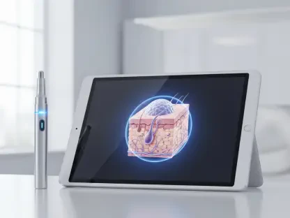

The possibility of a critically ill patient in the intensive care unit no longer needing to risk a dangerous transport to the radiology wing for life-saving metabolic imaging represents a profound shift in modern clinical practice. This transformation is currently spearheaded by a new portable positron emission tomography system developed by researchers at Washington University in St. Louis. By utilizing a flexible robotic-arm architecture, this innovative device eliminates the need for the massive, fixed installations that have long defined the field of nuclear medicine. This portability allows high-resolution functional imaging to be performed directly in intensive care units or operating rooms, places where patient stability is often too precarious for traditional travel. While bedside tools like ultrasound or CT are excellent for showing anatomy, they frequently fail to highlight the metabolic activity of a disease. This technology bridges that gap by providing functional insights in real-time, helping clinicians identify precisely where a disease is most active.
Overcoming the Logistical Hurdles of Traditional PET Scans
Standard PET/CT scanners have historically been confined to centralized laboratories due to their immense weight and the significant financial investment required for their installation and maintenance. This creates a substantial logistical bottleneck for patients who are too unstable to be transported across a large hospital campus. Moving these individuals increases the risk of serious medical complications, such as dislodged lines or respiratory distress, and delays urgent diagnostic decisions that are critical in acute care. The need for a mobile solution has become a top priority for emergency settings where time is the most valuable commodity. By reimagining the structural requirements of molecular imaging, the engineering team has managed to condense the capabilities of a room-sized machine into a maneuverable unit. This change addresses the fundamental conflict between the need for high-level metabolic data and the physical limitations of the most vulnerable patients in the medical system.
To address these structural challenges, the research team designed a prototype that replaces the traditional circular gantry with a flexible robotic arm and specialized detector panels. This design allows for what is known as arbitrary positioning, meaning the scanner can maneuver around bulky surgical equipment, ventilators, and other essential life-support systems found at the bedside. Clinicians can target specific organs or areas of interest without requiring the patient to be perfectly centered inside a restrictive and intimidating machine. Such flexibility is particularly ideal for crowded and sterile operating environments where space is at a premium and maintaining a clear field of motion is necessary. The robotic arm provides the degree of freedom required to capture data from multiple angles that were previously inaccessible. This mechanical innovation ensures that the diagnostic process adapts to the patient’s physical needs rather than forcing the patient to adapt to the machine’s constraints.
Faster Imaging with Incremental Reconstruction Technology
A major technical hurdle for bedside PET imaging has always been the excessive time required to process raw data into a viewable and diagnostic image for the medical team. In standard clinical practice, images are usually only available after a scan is entirely completed and the data has been sent to an external server for reconstruction. This delay is far too slow for active surgical procedures where immediate feedback can alter the course of an operation. The St. Louis team solved this problem by implementing an incremental algorithm that updates the image as new data is collected by the detectors. This allows for immediate visual feedback during the scanning process, enabling doctors to make decisions while the patient is still on the table. By processing data in small batches as the robotic arm moves, the system creates a dynamic workflow that mirrors the fast-paced nature of interventional medicine where every single minute matters for the patient outcome.
This workflow allows clinicians to watch a molecular map of the tissue develop in real-time as the scanning process unfolds before their eyes. As the robotic arm moves the detectors to different angles, the system feeds this new information into the existing image rather than starting the reconstruction from scratch. This adaptive approach ensures that the most relevant metabolic data is highlighted quickly, often revealing crucial structural details after only a few detector movements are completed. Experimental testing has shown that this real-time method maintains the same high-resolution quality found in conventional, stationary scanners. During studies using rod clusters to simulate complex human tissues, the system produced clear images that allowed researchers to make informed decisions long before a full scan was finished. This level of efficiency can drastically reduce procedural times and the duration of patient sedation, which significantly lowers the overall risk profile for high-risk clinical cases.
Redefining Oncology and Interventional Procedures
The arrival of portable PET technology supports a much broader trend toward decentralized and personalized healthcare in the modern medical landscape. By bringing high-level diagnostics directly to the point of care, hospitals can improve patient outcomes while simultaneously freeing up centralized radiology resources for routine outpatient scans. This concept of adaptive imaging allows operators to customize scans on the fly, focusing more intensely on specific areas of concern as they appear on the display screen. This level of customization was previously impossible with static machines that follow rigid, pre-programmed scanning paths regardless of the findings. By empowering physicians to adjust their diagnostic focus in the moment, the technology ensures that the specific clinical questions are answered with high precision. This evolution moves the imaging process from a passive observation to an active tool that clinicians can use to navigate complex biological landscapes in a variety of settings.
In the specialized field of oncology, this technology is a potential game-changer for delicate procedures like tumor biopsies and targeted ablations. Real-time PET guidance allows doctors to steer biopsy needles toward the most metabolically active hot spots within a tumor, ensuring the sample is diagnostically accurate and representative of the malignancy. For cancer treatments that use heat or cold to destroy tumors, the portable scanner provides exact boundaries of the malignancy during the procedure itself. This ensures the therapy is applied exactly where it is needed most, minimizing damage to surrounding healthy tissue. By integrating metabolic data with surgical precision, the system helps prevent the need for repeat procedures that can be taxing on a patient’s health. The ability to verify the success of a treatment before the patient even leaves the operating room could fundamentally alter how we approach local cancer therapies and long-term management strategies.
Future Implementation and Global Healthcare Impact
While currently functioning as a high-performance prototype, the journey toward a clinical-grade application is well underway with human trials projected to begin in 2027. The researchers are currently focusing on refining the system’s ergonomics and safety features to ensure it can handle the high-stakes environment of a real-world operating room or emergency department. These trials will be pivotal in determining how the system performs when it is fully integrated into daily clinical workflows alongside other medical personnel. Engineers are also looking for ways to streamline the user interface, making it intuitive enough for non-specialists to operate under the guidance of remote radiologists. Ensuring that the device is robust enough to withstand the rigors of hospital transport while maintaining delicate calibration is a primary focus of the current development phase. This preparation is essential for creating a tool that is not only scientifically advanced but also practically viable for busy hospital staff.
The potential for lower costs and a smaller physical footprint helped this device democratize access to molecular imaging for a wider range of medical facilities. By dismantling the physical and financial barriers associated with traditional PET/CT, this technology promised to integrate metabolic insights into the heart of interventional medicine. These advancements stood as a testament to the power of robotics and data science in improving patient-centered care. The researchers demonstrated that clinical mobility did not have to come at the expense of image quality or diagnostic depth. As the industry moved toward 2028 and beyond, the focus shifted toward establishing these portable systems as a standard component of every major intensive care unit. This transition offered a practical solution to the long-standing problem of imaging the most critically ill patients. It was clear that the successful implementation of such systems provided a new roadmap for the future of decentralized diagnostic tools in the healthcare sector.