
The long-standing medical reliance on invasive spinal taps and multi-million dollar imaging suites to decode the mysteries of the human brain is finally being challenged by a simple vial of saliva. For decades, the medical community operated under the assumption that the most sensitive data regarding neurological health remained locked behind the skull or within the spinal column. However, a significant technological leap has proven that the molecular indicators of complex disorders are circulating right under the tongue, waiting to be read by sophisticated sensors. This shift from traumatic “needle-in-the-spine” diagnostics to a non-invasive oral swab marks a pivotal moment where the most complex organ in the body finally becomes accessible through its most humble fluid.
The transition toward saliva-based testing represents more than just a convenience for the patient; it is a fundamental restructuring of neurological care. By capturing the molecular secrets of epilepsy, Parkinson’s disease, and schizophrenia in a liquid sample, researchers have effectively bridged the gap between advanced neuroscience and accessible clinical practice. This evolution suggests that the central nervous system is not an isolated fortress, but a dynamic system that constantly sheds light on its internal state through peripheral biofluids. As this technology matures, the fear associated with brain health monitoring begins to dissipate, replaced by a streamlined process that respects both biological complexity and patient comfort.
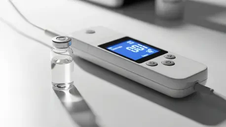
A Simple Spit Test Could Replace the Painful Lumbar Puncture
The days of relying on invasive spinal taps and multi-million dollar imaging suites to understand the human brain may be numbered. For a patient suffering from potential neurological decline, the diagnostic journey has historically been a gauntlet of anxiety and physical pain. Lumbar punctures require a long needle to be inserted between the vertebrae to withdraw cerebrospinal fluid, a process that carries risks of infection, chronic headaches, and significant discomfort. Even when successful, these procedures are often one-time events due to their intensity, leaving doctors with a single “snapshot” of a disease that is constantly evolving and changing.
A breakthrough study has revealed that the molecular secrets of epilepsy, Parkinson’s disease, and schizophrenia are not just hidden deep within the central nervous system—they are present in our saliva. This realization allows for a dramatic departure from traditional methods. Instead of high-risk procedures, a patient can now provide a sample through a simple, non-invasive oral swab that takes only seconds to collect. This shift democratizes brain health by making testing as easy as a routine dental check-up, removing the barriers that often prevent individuals from seeking early medical advice when symptoms first appear.
The accessibility of saliva also opens the door for longitudinal studies that were previously impossible. Because a spit test is painless and low-cost, medical professionals can collect samples weekly or even daily to observe how a patient responds to a new medication or how a condition fluctuates over time. This level of granularity in data collection provides a much clearer picture of the patient’s health than an occasional, invasive spinal tap ever could. It marks the end of an era defined by diagnostic trauma and the beginning of a more humane, data-driven approach to neurology.
The Critical Need for Non-Invasive Brain Health Monitoring
For decades, the blood-brain barrier has served as a formidable obstacle for doctors, trapping vital health markers within the brain and cerebrospinal fluid. This physiological wall is designed to protect the brain from toxins, but it also prevents the easy escape of disease indicators into the bloodstream. Consequently, many blood-based tests fail to provide the high-resolution data needed to distinguish between different types of neurological decline. Traditional diagnostic methods like Positron Emission Tomography (PET) scans are prohibitively expensive for routine screening and require specialized facilities that are not accessible to everyone, especially in rural or underserved areas.
Because these procedures are so taxing and costly, many neurological disorders are diagnosed only after symptoms become severe, missing the critical window for early intervention. This technological gap has created an urgent demand for a diagnostic tool that is both affordable and patient-friendly. When a diagnosis is delayed until physical tremors or cognitive lapses are unmistakable, the underlying damage to the neural pathways is often extensive. A non-invasive monitoring system allows for a proactive rather than reactive stance, enabling clinicians to identify chemical imbalances before they manifest as life-altering symptoms.
Furthermore, the lack of frequent monitoring tools has hindered the development of personalized treatment plans. Every brain is unique, and the way a specific individual metabolizes a drug or progresses through a disease varies wildly. Without a low-barrier way to check brain markers regularly, doctors are often forced to rely on trial-and-error prescribing. An affordable, saliva-based diagnostic platform bridges this gap, providing the frequent feedback loop necessary to refine treatments in real-time. This urgency for change has driven the scientific community to look beyond traditional methods toward more innovative, light-based detection systems.
The Science of Sound and Light: How GME-SERS Detects Disease
Traditional tests often fail because they only look at the amount of protein in a sample, which fluctuates naturally based on diet, stress, or hydration. This new method focuses on “conformational dynamics”—the specific way proteins fold and change shape as a disease progresses. In many neurological conditions, proteins do not just increase in volume; they begin to misfold and clump together, creating toxic structures. By analyzing the structural integrity of these proteins rather than their raw concentration, the test provides a much more stable and reliable indicator of a person’s neurological state.
Using a specialized nanostructure of copper oxide and gold (Au–CuO), researchers created “hotspots” that amplify weak molecular signals by over a billion times. This technology, known as Surface-Enhanced Raman Scattering (SERS), works by bouncing laser light off the molecules in the saliva. Normally, the signals produced by neuroproteins are so faint that they are lost in the background noise of the fluid. However, the unique properties of the Au–CuO nanostructure act like a molecular megaphone, catching these faint vibrations and magnifying them until they can be clearly identified as a specific chemical signature.
The test identifies the exact moment when healthy protein monomers transform into toxic fibrils. These structural changes act as a molecular “fingerprint” unique to specific neurological conditions like Parkinson’s or schizophrenia. Unlike other laboratory tests that require chemical dyes or radioactive tracers, this Raman-based technology analyzes samples in their natural state. This “label-free” approach ensures high purity and reduces the complexity of the diagnostic process, making the results easier to interpret and less prone to interference from external reagents.
Proven Precision: Clinical Validation and High Accuracy Rates
The efficacy of this technology was put to the test in a collaborative study with St. Vincent’s Hospital, involving a diverse group of patients and healthy controls. The researchers sought to prove that the GME-SERS platform could function in a messy, real-world clinical environment rather than just a controlled laboratory setting. By testing individuals with confirmed cases of epilepsy, schizophrenia, and Parkinson’s disease against a control group of healthy volunteers, the team established a rigorous benchmark for the sensor’s performance. The results confirmed that the saliva-based platform is a clinical powerhouse capable of handling the complexities of human biology.
The platform demonstrated a classification accuracy exceeding 90% across the board, with some tests reaching a staggering 98% precision in identifying specific disorders. This level of accuracy is nearly unprecedented for a non-invasive test, placing it on par with, or even exceeding, the reliability of much more expensive imaging techniques. The ability to achieve such high precision from a single drop of saliva proved that the “fingerprinting” technique was successfully capturing the unique structural signatures of diseased proteins. This validation demonstrated that the technology could effectively filter out the “noise” of other substances found in the mouth to find the critical markers of brain health.
The technology proved capable of differentiating between epilepsy, schizophrenia, and Parkinson’s disease based solely on the structural signatures found in saliva. This is a critical distinction, as many neurological symptoms can overlap, making a definitive diagnosis difficult for even the most experienced neurologists. The research has gained international recognition, with findings published in the prestigious journal Advanced Materials, highlighting its potential to set a new global standard in medical materials science. Peer validation from the global scientific community has solidified the platform’s reputation as a legitimate and revolutionary tool for the future of medicine.
From the Lab to the Clinic: Practical Applications and Future Implementation
The transition of this technology into everyday medical practice promises to democratize neurological care through several key strategies. Researchers are currently working to miniaturize the Raman sensors, moving them from large laboratory setups into portable, handheld devices for use in local clinics. This evolution is essential for making the technology truly accessible, as it removes the need for a central laboratory and allows for immediate results. A doctor in a small community clinic could theoretically perform the test and provide a diagnosis during a single office visit, significantly reducing the time and travel burden on the patient.
Because the test is non-invasive and low-cost, patients with schizophrenia or Parkinson’s can be tested frequently to monitor disease progression and the effectiveness of their medications. This represents a massive shift in chronic disease management. Instead of waiting months between appointments to see if a medication is working, clinicians can use the saliva test to see molecular changes within days. This rapid feedback allows for much more precise dosage adjustments, potentially reducing the side effects of powerful neurological drugs and improving the overall quality of life for the patient.
The affordability of the GME-SERS platform allows for the implementation of routine neurological screenings during annual physical exams, potentially catching brain disorders years before physical symptoms appear. Much like a glucose monitor for diabetics, the ultimate goal is to provide patients with the tools to monitor their own brain health at home, reducing the burden on the healthcare system. This proactive model of care could lead to a future where neurological diseases are managed as manageable chronic conditions rather than unexpected, devastating crises.
The research team successfully demonstrated that nanostructured materials could bridge the gap between complex brain pathology and simple clinical diagnostics. By achieving high accuracy through a non-invasive medium, they provided a viable alternative to the traditional, painful methods of the past. The collaborative effort between materials scientists and medical professionals ensured that the technology remained grounded in clinical reality while pushing the boundaries of physics. This initiative proved that the structural fingerprints of disease were accessible through saliva, offering a new pathway for early detection and personalized treatment. The focus moved toward the engineering of scalable, consumer-ready devices that promised to make brain health monitoring a routine part of modern life.


