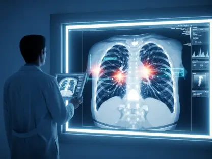

The historical reliance on traditional monoaminergic antidepressants has left a significant portion of the psychiatric patient population in a state of perpetual search for relief, particularly those suffering from treatment-resistant depression. While standard selective serotonin reuptake inhibitors typically require several weeks or even months to manifest therapeutic benefits, they fail to provide any meaningful remission for approximately one-third of diagnosed individuals. This clinical stagnation has necessitated a shift toward rapid-acting interventions, with ketamine emerging as a frontrunner in modern neuropharmacology. Despite its proven efficacy in clinical settings, the specific molecular mechanisms by which ketamine reorganizes the human neural architecture remained largely theoretical for years. However, a landmark study conducted by researchers at Yokohama City University has finally moved beyond animal models and indirect imaging to provide a direct visualization of how this substance reshapes brain chemistry. By focusing on the immediate physical changes within the living human brain, scientists are now able to explain why ketamine succeeds where conventional pills have consistently failed, marking a pivotal moment in the transition toward biologically grounded mental healthcare.
Visualizing Molecular Changes with PET Technology
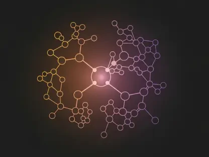
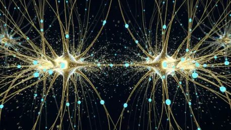
The scientific community has long suspected that the primary driver of ketamine’s rapid antidepressant effect is the modulation of glutamate $\alpha$-amino-3-hydroxy-5-methyl-4-isoxazole propionic acid receptors, commonly referred to as AMPARs. These receptors are fundamental to synaptic plasticity, acting as the gatekeepers for communication between neurons and enabling the brain to adapt to new information or emotional states. In previous years, verifying the movement and density of these receptors in living humans was nearly impossible due to the limitations of standard imaging. While Functional Magnetic Resonance Imaging can track changes in blood flow and oxygenation, it lacks the molecular resolution required to see specific receptor proteins at the synaptic level. To solve this, the research team utilized a cutting-edge positron emission tomography tracer known as [11C]K-2. This specialized radioactive ligand was engineered to bind specifically to cell-surface AMPARs, allowing the researchers to generate high-resolution, three-dimensional maps of receptor distribution across the entire brain of patients suffering from severe, treatment-resistant depression.
The methodology employed in this study involved a rigorous comparison between 34 patients with treatment-resistant depression and a control group of 49 healthy individuals to establish a baseline for receptor density. Over a fourteen-day trial period starting in early 2026, the patients received either intravenous ketamine or a saline placebo, with PET scans performed at strategic intervals to capture the shifting molecular landscape. The use of the [11C]K-2 tracer allowed the team to observe, for the first time, that depression is not merely a “chemical imbalance” of neurotransmitters like serotonin but is deeply rooted in the physical density and availability of these glutamate receptors. By mapping these receptors before and after treatment, the study provided definitive proof that ketamine’s primary function is to trigger a rapid redistribution of the brain’s communication hardware. This technological leap from indirect observation to direct molecular visualization effectively bridges the gap between laboratory neuroscience and clinical psychiatry, providing a clear biological framework for understanding how rapid-acting antidepressants function in a real-world medical context.
Rebalancing the Brain’s Reward and Function Centers
The data gathered from the PET scans revealed that ketamine does not affect the brain uniformly; instead, it initiates a highly localized and strategic rebalancing of neural circuitry. In patients with chronic depression, the researchers identified significant receptor abnormalities in areas responsible for both executive function and emotional processing. Upon the administration of ketamine, a notable increase in AMPAR density occurred within the cortical regions, particularly those areas involved in complex decision-making and the regulation of mood. This “upregulation” of receptors effectively strengthens the neural pathways that allow an individual to engage with the world and manage their emotional responses. By increasing the density of these receptors in the cortex, ketamine restores the brain’s ability to process positive information and maintain cognitive flexibility, which are often the first faculties lost during a major depressive episode. This physical restoration of the cortex provides the structural foundation for the cognitive clarity many patients report shortly after their first treatment session.
Equally important to the increase in cortical receptors was the simultaneous decrease in receptor density observed in the habenula, a small but powerful structure located deep within the brain. The habenula is often described by neuroscientists as the “anti-reward” center because it becomes hyperactive in response to disappointment, pain, and perceived failure. In many depressed individuals, this region is stuck in an overactive state, constantly signaling a sense of despair and inhibiting the brain’s natural reward systems. The PET imaging showed that ketamine effectively “muffles” these signals by reducing the concentration of AMPARs in the habenula. This dual action—strengthening the higher-order thinking centers while silencing the biological sources of pessimism—creates a window of opportunity for patients to escape the cyclical nature of depressive thought patterns. This regional specificity explains why ketamine is so uniquely effective; it doesn’t just provide a general boost to mood but physically recalibrates the balance between the brain’s systems for resilience and its systems for processing emotional pain.
Linking Physical Changes to Patient Recovery
Perhaps the most groundbreaking aspect of the Yokohama City University study was the discovery of a nearly perfect correlation between the scale of physical receptor changes and the degree of clinical improvement reported by patients. Using standardized depression rating scales, the researchers found that individuals who experienced the most significant redistribution of AMPARs also saw the most dramatic reduction in their symptoms. This finding is revolutionary because it provides an objective, biological metric for a condition that has historically been diagnosed based on subjective self-reporting and behavioral observation. The fact that the physical “reshaping” of the brain directly mirrors the psychological recovery suggests that these molecular shifts are not merely a side effect of the drug, but are the actual mechanism of healing. This evidence moves psychiatry closer to the standards of internal medicine, where treatment success can be measured through tangible biological markers rather than just a patient’s perceived improvement in their daily mood or energy levels.
The ability to link specific molecular movements to clinical outcomes paves the way for a more personalized, biomarker-driven approach to mental healthcare. In the current landscape, many patients spend years cycling through various medications in a process of trial and error that can be both exhausting and dangerous. However, if PET imaging can be used to identify specific receptor imbalances at the start of treatment, clinicians could theoretically predict which patients are most likely to respond to ketamine before the drug is even administered. This would allow for a level of precision medicine previously unheard of in psychiatry, where the biological profile of a patient’s brain dictates the therapeutic strategy. While the study confirms that ketamine is a powerful tool for many, it also highlights that those who do not show specific receptor changes may require different interventions. This nuanced understanding ensures that medical resources are directed toward the most effective treatments for each individual, reducing the time spent on ineffective therapies and accelerating the path to long-term mental stability.
Addressing Research Limitations and Future Applications
While the visual evidence of ketamine’s impact is compelling, the transition from a controlled research study to widespread clinical practice faces several logistical and financial hurdles that must be addressed in the coming years. The current study focused on a fourteen-day window, which provided excellent insights into the acute phase of treatment but left questions regarding the long-term durability of these molecular changes. It is not yet clear whether the increased receptor density in the cortex remains stable over several months or if the brain eventually reverts to its previous depressed state without maintenance doses. Future longitudinal studies will need to track patients over longer periods to determine the optimal frequency of ketamine administration required to sustain these physical brain changes. Furthermore, the specialized [11C]K-2 PET tracer is currently expensive and requires sophisticated laboratory equipment that is not available in standard hospitals, meaning that using these scans as a routine diagnostic tool remains a future goal rather than a current reality for most.
Building on these findings, the next logical step for the medical community involves the development of more accessible ways to monitor or stimulate these receptor changes. While PET scans are the gold standard for visualization, researchers are already looking into whether more affordable blood-based biomarkers or specific EEG patterns can serve as proxies for the receptor density changes seen in this study. Additionally, the discovery that the habenula and the cortex are the primary sites of action for ketamine allows pharmaceutical companies to design new, even more targeted molecules that can influence these specific regions with fewer side effects. The objective should be to move toward a model where every patient with treatment-resistant depression receives a personalized “brain map” that guides their therapy. By prioritizing the integration of molecular imaging with clinical practice, the psychiatric field can finally move away from the “one size fits all” approach and toward a future where the physical reshaping of the brain is a standard, measurable, and highly successful part of mental health recovery.