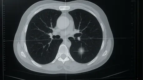

The high risk of overfitting in medical machine learning is mitigated through rigorous variance screening and Pearson correlation to remove redundant imaging data. This technical principle is now at the forefront of efforts to resolve one of the most persistent dilemmas in thoracic oncology: the management of ground-glass nodules (GGNs) detected on chest CT scans. These nodules, which appear as faint, hazy clouds within the lungs, represent a diagnostic “gray zone” where the human eye often fails to distinguish between benign inflammation, pre-cancerous growths, and early-stage invasive cancers. Because GGNs do not fully obscure the underlying structures of the lung, such as blood vessels and bronchial walls, radiologists face significant uncertainty when determining the level of aggression required for treatment. For decades, this ambiguity has forced clinicians into a strategy of watchful waiting, a period defined by recurring scans and significant patient anxiety. The risk remains that during this monitoring phase, a slow-growing nodule might transition into an invasive adenocarcinoma before a surgical decision is made. Consequently, there is an urgent need for more objective, data-driven methodologies that can look beyond the visual surface of these smudges to reveal their true biological potential and provide a clear roadmap for clinical intervention.
The Technological Shift: Turning Pixels into Biological Data
Radiomics represents a fundamental evolution in diagnostic imaging by treating standard medical scans as high-dimensional quantitative data rather than just visual representations. This field involves the high-throughput extraction of thousands of features from digital images, including descriptors of shape, size, intensity distribution, and texture. By utilizing mathematical algorithms to analyze the spatial relationships between pixels, radiomics can uncover complex patterns that are entirely invisible to the most experienced human observers. The underlying hypothesis of this discipline is that these “hidden” imaging phenotypes are directly linked to the genomic and proteomic characteristics of the tissue. In the context of pulmonary care, this allows a standard CT scan to serve as a non-invasive window into the cellular architecture of a nodule. As these analytical tools become more sophisticated, the focus is shifting toward creating standardized numerical scores that can reliably predict the aggressiveness of a lesion, thereby moving the diagnostic process away from qualitative descriptions and toward a more reproducible, objective framework that enhances clinical confidence.
A recent study conducted in Foshan, China, provided a compelling demonstration of this technology’s capabilities by analyzing a cohort of 253 patients presenting with ground-glass nodules. All participants in this research underwent surgical resection, which allowed the team to use the definitive pathological diagnosis as the “gold standard” for evaluating the AI’s predictions. The researchers focused on the entire spectrum of pulmonary adenocarcinoma, categorizing the lesions into four critical stages: atypical adenomatous hyperplasia (AAH), adenocarcinoma in situ (AIS), minimally invasive adenocarcinoma (MIA), and invasive adenocarcinoma (IAC). Of the 253 cases examined, the majority were identified as fully invasive, while the remainder represented various pre-cancerous or early-stage growths. By analyzing this wide spectrum, the study sought to determine whether specific radiomic signatures could accurately map the transition of a nodule from a localized, low-risk proliferation of cells into a dangerous malignancy. This comprehensive approach is essential for developing a tool that can be used in real-world clinical environments where nodules exist at various points in their biological evolution.
Methodological Precision: From Nodule Segmentation to Model Building
The technical workflow of the study was meticulously structured to ensure that the resulting predictive model was both stable and accurate. The process began with 3D segmentation, where radiologists manually outlined the boundaries of each nodule across several CT slices to create a volumetric “Region of Interest.” From these 3D volumes, a total of 1,836 distinct radiomic features were extracted, covering first-order statistics, shape-based metrics, and advanced texture descriptors such as the Gray Level Co-occurrence Matrix. This high-throughput extraction provided a detailed mathematical fingerprint of each lesion, capturing nuances in density and structural complexity that traditional measurements often ignore. By prioritizing 3D volumetric analysis over traditional 2D measurements, the researchers ensured that the entire mass of the nodule was accounted for, which is critical since invasive components often develop asymmetrically within the lesion. This level of detail allows for a much more nuanced understanding of the nodule’s internal architecture and its likely behavior in the future.
To translate this massive volume of data into a practical diagnostic tool, the researchers employed a multi-stage dimensionality reduction process to prevent model overfitting and ensure generalizability. Since the number of features extracted far exceeded the number of patients, it was necessary to filter out redundant and irrelevant data points. The team utilized techniques such as LASSO regression to identify a small subset of “master” features that possessed the strongest correlation with the actual pathological results. These final features were then used to train a Random Forest classifier, a robust machine learning algorithm that aggregates the outputs of multiple decision trees to produce a highly stable prediction. This rigorous selection process ensured that the model was not merely memorizing the noise present in the specific dataset but was instead identifying the fundamental imaging characteristics that define different stages of cancer. By refining the data down to its most potent elements, the researchers created a model that is more likely to remain accurate when applied to new, unseen patient populations in different healthcare settings.
Clinical Stratification: Identifying the Invasive Threshold
The researchers organized their findings into three distinct classification tasks that were specifically designed to mirror the clinical decisions faced by thoracic surgeons. Task 1 aimed to distinguish pre-cancerous lesions from all other nodules, while Task 2 focused on separating non-invasive stages from invasive disease. The third and most critical task was the identification of fully invasive adenocarcinoma (IAC) versus all other categories. This distinction is vital because the diagnosis of IAC often dictates a more aggressive surgical approach, such as a lobectomy, to ensure that the cancer does not recur or spread. In contrast, earlier stages like AIS or MIA might only require a more conservative wedge resection, which preserves more of the patient’s lung function and improves their postoperative quality of life. By focusing on these specific milestones, the radiomics model provides a quantitative basis for selecting the most appropriate surgical intervention, potentially reducing the physical burden on patients while maintaining high standards of oncological safety.
The performance of the model in these tasks was highly impressive, particularly regarding the identification of fully invasive cancer. In Task 3, the radiomics system achieved an Area Under the Receiver Operating Characteristic Curve (AUC) of 0.9484, demonstrating a high degree of predictive accuracy. Furthermore, the model maintained a specificity of 0.9500, which is a critical metric for any diagnostic tool intended for clinical use. High specificity means that the AI was exceptionally good at avoiding “false alarms,” ensuring that patients with low-risk or non-invasive nodules were not mistakenly identified as having aggressive cancer. This precision is essential for preventing the overtreatment of patients and ensuring that intensive surgical resources are reserved for those who truly need them. These results suggest that radiomics can offer a level of diagnostic clarity that significantly exceeds visual inspection alone, providing clinicians with a reliable, objective metric to support their decision-making process in the management of complex lung nodules.
Comparative Analysis: Radiomics Versus Deep Learning Frameworks
A unique aspect of this research was the comparison between traditional radiomics and deep learning models using a ResNet18 neural network. While deep learning is often celebrated for its ability to automatically discover relevant patterns from images, it did not outperform the radiomics approach in this specific study. The deep learning models achieved AUC scores between 0.82 and 0.85, which were notably lower than the results produced by the handcrafted radiomics model. This discrepancy was likely due to the type of data used; the radiomics approach utilized high-fidelity raw imaging data from the CT volumes, whereas the deep learning models were restricted to standard display images. This finding highlights the critical importance of data quality and feature engineering in medical AI. It suggests that for the specific and highly nuanced task of GGN stratification, the structured extraction of mathematically defined features remains the more robust methodology, providing a more reliable foundation for clinical decisions than a “black box” deep learning system.
This comparison also suggests that the future of medical imaging may lie in a hybrid approach that combines the strengths of various machine learning methodologies. While deep learning is unparalleled for tasks like automatic nodule detection and segmentation, radiomics provides a more precise and interpretable pathological assessment. By integrating these two technologies, developers can create more efficient diagnostic platforms that automate the initial detection of nodules while providing deep, quantitative insights into their biological nature. Such an integrated system would allow radiologists to process larger volumes of cases with higher accuracy, focusing their expertise on the most complex scenarios while the machine handles the routine data extraction. This synergy between human-defined features and machine-discovered patterns could lead to a more comprehensive diagnostic toolset, ultimately improving the speed and precision with which lung cancer is diagnosed and treated in clinics around the world.
Future Pathways: Validating the Virtual Biopsy in Clinical Practice
Despite the success of the current study, several challenges must be addressed before radiomics can be implemented as a standard of care. The research was a single-center, retrospective study, which means the model was developed and tested on data from a specific hospital with standardized equipment and protocols. For these tools to be globally applicable, they must be validated through multi-center trials involving diverse patient populations and a variety of CT scanners from different manufacturers. This validation is necessary to ensure that the radiomic signatures remain consistent across different imaging settings and that the model’s accuracy does not degrade when faced with variations in scan quality or radiation dosage. Additionally, the researchers identified the need to test these models in a broader screening environment, where the prevalence of benign nodules is higher, to ensure that the system remains effective in identifying the early signs of malignancy within a larger, more varied pool of cases.
The study from Foshan successfully established the concept of the “virtual biopsy” as a viable method for managing the complexities of lung adenocarcinoma. By converting visual patterns into quantifiable data, the research demonstrated that machine learning could effectively navigate the adenocarcinoma spectrum and provide a non-invasive glimpse into a tumor’s invasive potential. This project showed that the integration of radiomics into clinical workflows could significantly reduce the subjectivity of nodule interpretation and foster a more personalized approach to surgery. Clinicians were encouraged to adopt these tools as diagnostic adjuncts that could clarify ambiguous findings and help prioritize high-risk patients for immediate treatment. As these models continued to undergo rigorous prospective validation, the prospect of replacing “watchful waiting” with a decisive, data-driven strategy became increasingly realistic. The transition toward this more objective model of oncology offered a promising pathway for improving patient outcomes and streamlining the delivery of modern thoracic care.