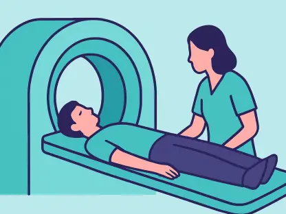

The global healthcare landscape is currently grappling with an annual toll of approximately 250,000 lives lost to brain tumors, a statistic that underscores the critical need for earlier and more precise diagnostic interventions. While magnetic resonance imaging remains the gold standard for visualizing these neurological threats, the sheer volume of data generated daily places an immense cognitive burden on human radiologists. This workload can occasionally lead to fatigue-induced errors or delays that are unacceptable in high-stakes oncology. In response, researchers at the University of Sharjah have pioneered a sophisticated artificial intelligence system that does not merely automate detection but refines the entire diagnostic process. By integrating advanced machine learning architectures, this technology promises to bridge the gap between initial screening and specialized treatment planning, providing a rapid second opinion that maintains the highest standards of clinical accuracy.
A Strategic Two-Stage Diagnostic Framework
Step 1: Mirroring the Clinical Radiologist Workflow
The foundation of this diagnostic innovation lies in its structured, two-tier approach, which intentionally separates the initial detection of an abnormality from its subsequent classification. Traditional medical AI often attempts to solve multiple diagnostic problems simultaneously, which can lead to increased computational overhead and a higher probability of false positives. By contrast, the University of Sharjah’s framework initiates the process with a binary check, determining solely whether a tumor is present or absent within the MRI scan. If the system finds no evidence of pathology, it halts the analysis, thereby preserving computational resources and allowing the specialist to focus on other cases. This streamlined logic mirrors the triage process used in busy emergency departments, where the primary goal is to quickly identify patients who require immediate attention. By filtering out healthy scans, the system ensures that more complex tools are reserved for confirmed cases.
Step 2: Determining Pathological Specificity
When the initial screening phase confirms the presence of a growth, the system transitions into a sophisticated multi-class diagnostic phase to determine the specific nature of the tumor. This stage is critical because the medical management of a glioma, which is often aggressive and infiltrating, differs fundamentally from the treatment of a meningioma or a pituitary tumor. The AI must distinguish between these types by analyzing subtle textural variations and structural patterns that might be overlooked by the human eye during a routine review. By providing this detailed categorization immediately following detection, the technology eliminates the need for secondary manual reviews that often delay the start of therapy. This sequential methodology ensures that the diagnosis is not just fast but also contextually relevant to the specific oncological challenge at hand. It creates a robust pipeline where the final output is a comprehensive profile that serves as a definitive starting point.
Architectural Synergy and Data Integrity
Technology: Optimizing CNNs, LSTMs, and Transformers
Achieving the required level of precision in neuro-oncology necessitates an architectural synergy that combines the strengths of various deep learning models. While Convolutional Neural Networks have long been the industry standard for processing visual information, the Sharjah research team enhanced their performance by integrating Long Short-Term Memory layers and attention mechanisms. This hybrid design allows the model to retain critical spatial information while simultaneously focusing on the most relevant features of the MRI image, effectively ignoring the visual noise that often plagues medical scans. Furthermore, the introduction of the Vision Transformer has revolutionized the initial detection phase by enabling a more holistic view of the brain’s anatomy. Unlike older models that process images in localized patches, the Vision Transformer utilizes self-attention to capture global context. This ensures the AI can detect large-scale structural deviations that might be missed if it were too narrowly focused on small pixel groups.
Data Quality: Establishing Consistency With Preprocessing
The reliability of any artificial intelligence model is deeply tethered to the quality and consistency of the data it processes, particularly in the diverse world of medical imaging. For this reason, the researchers utilized a massive dataset of over 7,000 MRI images, subjecting each one to a rigorous preprocessing pipeline before it reached the neural networks. This stage is vital because MRI scans often vary significantly depending on the manufacturer of the scanner, the specific hospital settings, and the orientation of the patient. By standardizing every image for size, brightness, and contrast, the team removed technical artifacts that could lead to algorithmic bias. This normalization process ensures that the AI makes its decisions based on the actual biological reality of the tumor rather than the technical quirks of the imaging hardware. Consequently, the system demonstrates high robustness, maintaining its accuracy across different clinical environments and protocols, which is key for broad adoption.
Practical Implementation and the Road Ahead
Efficiency: Maximizing Speed and Personalized Outcomes
Efficiency is a paramount concern in modern hospital settings where every minute can influence the success of a surgical intervention or radiation therapy. The AI system developed by the University of Sharjah demonstrated remarkable performance in this regard, achieving inference times as low as 20 milliseconds per scan. This near-instantaneous processing capability allows for the integration of the tool directly into the radiologist’s workstation, providing real-time validation of their findings without adding to their administrative burden. Beyond the speed of diagnosis, the precision of the classification stage directly impacts the shift toward personalized medicine. When a doctor can immediately confirm whether a growth is a benign meningioma or a malignant glioma, they can bypass general observation periods and move straight to targeted treatment plans. This acceleration of the care pathway not only improves physiological outcomes but also reduces the psychological stress associated with long waiting periods.
Reliability: Ensuring Model Robustness and Interpretability
Despite the significant advancements represented by this technology, several practical hurdles must be cleared before it becomes a ubiquitous feature in global healthcare systems. Future initiatives will likely focus on expanding the diversity of training datasets to include rarer tumor types and images from lower-quality scanners found in developing regions. Ensuring that the AI remains accurate regardless of the equipment used is essential for promoting health equity. Additionally, the development of explainability tools is a critical next step in fostering trust between the machine and the clinician. By creating heat maps that explain why the AI identified a certain region as a tumor, the system moves away from being an opaque black box and becomes a transparent partner in the diagnostic process. This interpretability will allow doctors to verify the AI’s logic, ensuring that the final medical decision remains in human hands while being supported by the most advanced computational tools currently available to them.
Strategic Integration: Moving Toward Automated Neuro-Radiology
The successful development of this two-stage AI framework provided a clear blueprint for the future of automated neuro-radiology. Medical institutions that prioritized the integration of these tools into their standard workflows observed a marked improvement in diagnostic throughput and a reduction in missed pathologies. To fully capitalize on these gains, healthcare providers were encouraged to invest in the digital infrastructure necessary to support high-speed AI inference and to establish clear protocols for human-AI collaboration. The focus moved from simply proving that AI can detect tumors to ensuring that it can do so within the messy, heterogeneous environment of a functioning hospital. Ultimately, the transition to these intelligent systems offered a pathway toward a more proactive and precise form of oncology, where early detection became the norm rather than the exception. By embracing these architectural synergies, the medical community took a significant step toward neutralizing the threat of tumors through power of analysis.