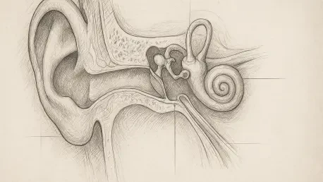

Traditional diagnostic methods for middle ear disorders have historically forced surgeons to rely on audiometric patterns and invasive exploratory tympanotomy procedures. This necessity often placed patients in a precarious position, as the precise nature of their hearing deficit remained unknown until a surgical incision was physically made. In 2026, the landscape of otology is shifting toward a more transparent diagnostic model where the hidden structures of the middle ear are visualized with surgical precision. The ossicular chain, comprising the malleus, incus, and stapes, represents a delicate mechanical bridge that must function perfectly to ensure clear hearing. When these bones are malformed or fused, sound cannot effectively reach the inner ear, leading to significant conductive loss. Recent clinical evaluations from major medical institutions have focused on using high-resolution computed tomography paired with three-dimensional reconstruction to solve this anatomical puzzle. By integrating these scans, the medical community is finding new ways to improve the accuracy of diagnoses and the success rates of delicate ear operations.
Precision Through Advanced Digital Reconstruction
The transition from standard two-dimensional “slices” to three-dimensional rendering represents a major leap in otological diagnostics. While traditional CT scans provide important data, they require surgeons to mentally piece together flat images to understand the complex three-dimensional reality of the ear’s anatomy. This process is inherently subjective and prone to human error, especially when dealing with the smallest bones in the human body. By utilizing advanced post-processing techniques, clinicians can now generate a holistic view of the middle ear cavity, allowing for a thorough inspection of the bones from every possible angle. This digital evolution is not merely about aesthetic clarity; it is about providing a functional map that reflects the actual physical constraints the surgeon will face. As these imaging protocols become more standardized in 2026, the reliance on guesswork is being replaced by a sophisticated, data-driven approach that prioritizes patient safety and anatomical accuracy above all else.
Enhancing Surgical Planning: 3D Models
By using multiplanar reformation, clinicians can rotate and view the middle ear bones as discrete objects, allowing them to spot tiny malformations or displacements that might be invisible on a standard scan. This level of detail is essential for identifying congenital issues, such as a missing stirrup leg or fused joints, ensuring the surgeon is fully prepared before entering the operating room. The ability to see these structures in a virtual, high-fidelity environment allows for a trial-run of the surgical approach, where the medical team can evaluate the most effective way to navigate the narrow confines of the temporal bone. This preoperative visualization acts as a critical rehearsal, highlighting potential obstacles like an unusually positioned facial nerve or an anomalous bony growth that could complicate the procedure. Consequently, the surgical team can enter the theater with a refined strategy, having already identified the optimal path to reach the site of the pathology, which significantly reduces the time the patient remains under general anesthesia.
Tactical Advantages: Surgeon-Led Imaging
The value of this technology is further amplified when the reconstruction is guided by the surgeon rather than a radiologist alone. Because the treating physician understands the specific clinical questions at hand, they can tailor the digital model to highlight critical areas, such as the proximity of the facial nerve or the exact shape of a malformed bone. This collaborative approach to imaging ensures that the surgical plan is highly personalized and technically grounded. When a surgeon performs the 3D post-processing, they are essentially performing a digital dissection, gaining insights into the unique anatomy of the patient that a standard report might miss. This hands-on involvement allows for the selection of the right prostheses and the anticipation of anatomical hurdles in advance, which streamlines the entire operative workflow. By the time the first incision is made, the surgeon has already navigated the patient’s anatomy hundreds of times in a digital space, leading to a much higher level of technical precision and a notable reduction in the risk of unexpected complications during the actual operation.
Analyzing Clinical Outcomes and Diagnostic Success
The implementation of 3D CT imaging has shown an impressive ability to match real-world surgical findings, bridging the gap between digital prediction and biological reality. In clinical studies throughout 2026, there has been a near-perfect agreement rate between the preoperative 3D diagnoses and what surgeons eventually discover during exploratory tympanotomy. This high degree of accuracy spans various conditions, including congenital ossicular anomalies, which are the most common cause of hearing loss in these patients, as well as cases of otosclerosis and traumatic breaks in the bone chain. Having this information beforehand removes much of the anxiety historically associated with middle ear surgery for both the patient and the provider. The diagnostic clarity provided by these scans allows for a more honest and detailed conversation regarding the expected outcomes and risks of the procedure. As the medical community continues to refine these imaging algorithms, the goal is to make the “exploratory” part of the surgery entirely obsolete, moving toward a model where every intervention is a planned correction.
Diagnostic Reliability: Identifying Structural Defects
The consistency of these scans is particularly evident when dealing with congenital malformations, which can be highly variable and unique to each individual. In a detailed review of patient data, researchers noted that 3D reconstructions correctly identified the status of the ossicular chain in nearly 97 percent of cases. Whether the hammer was fused to the anvil or the stirrup was completely missing its posterior crus, the digital models provided an accurate blueprint that was later confirmed in the operating room. This diagnostic reliability is a game-changer for pediatric cases, where congenital defects are more prevalent and surgical precision is even more vital due to the smaller size of the structures involved. By having a confirmed diagnosis before surgery, the medical team can ensure that all necessary specialized equipment and various sizes of prostheses are readily available. This logistical preparation, fueled by accurate imaging, prevents delays and ensures that the corrective procedure is completed as efficiently as possible, maximizing the chances of a successful long-term hearing restoration for the patient.
Technology Constraints: The Soft Tissue Challenge
Despite these successes, it is important to recognize that imaging technology has specific limitations, particularly regarding soft tissue and early-stage disease. While 3D CT scans are excellent for mapping bone, they can struggle to detect thin fibrous connections or early-stage tissue changes that have a similar density to surrounding fluids or mucosal linings. This “blind spot” means that while the scans provide a superior map of the skeletal structure, surgeons must still rely on their clinical judgment and audiometric data to fill in the gaps. For instance, a thin bridge of tissue connecting two bones might provide some mechanical stability that is not captured on the scan, potentially leading to a slight discrepancy in the predicted mobility of the chain. This limitation highlights the necessity of maintaining traditional surgical skills and diagnostic rigor alongside the new technology. An integrated approach—combining the power of the 3D scan with traditional tools like tuning fork tests and acoustic immittance—remains the most effective way to ensure a comprehensive understanding of the patient’s condition.
Measuring the Impact on Patient Hearing Recovery
The ultimate benchmark for any surgical innovation is the improvement in the patient’s quality of life, and the results following 3D-guided ear surgery are highly encouraging. Patients have shown massive leaps in their ability to hear daily sounds, with average hearing thresholds improving significantly after their bones were reconstructed using these digital maps. In the medical field, the “air-bone gap” is a key metric for success; reducing this gap to less than 20 decibels is the gold standard, a goal achieved by the vast majority of patients who underwent these image-guided procedures. This restoration of sound conduction does more than just increase volume; it restores the natural amplification system of the human body, allowing for better speech recognition and sound localization. For many individuals, this means a return to normal social activities and a significant reduction in the cognitive load associated with struggling to hear. The precision of the 3D-guided approach ensures that the prosthetic reconstruction mimics the original anatomy as closely as possible, leading to a more natural and sustainable hearing outcome.
Quantitative Success: Restoring Sound Pathways
The specific data regarding hearing gains highlights the transformative potential of this technology-driven surgical approach. Clinical results from 2026 demonstrated that the mean air-conduction thresholds improved by over 30 decibels following reconstruction, which represents a profound change in auditory perception. Most patients moved from a state of moderate-to-severe hearing loss to a level that no longer requires the use of external hearing aids for daily communication. This success is directly tied to the surgeon’s ability to use the 3D CT map to choose the perfect length and angle for the ossicular prosthesis. When the mechanical bridge is restored with such high accuracy, the transfer of energy from the eardrum to the inner ear is maximized, minimizing the loss of sound pressure. The high rate of patients reaching the benchmark air-bone gap proves that the integration of 3D imaging into the surgical workflow is not just a technical luxury but a clinical necessity for achieving the best possible results. These quantitative metrics provide a clear evidence base for the wider adoption of 3D reconstruction in otology centers worldwide.
Physiological Optimization: Improving Bone Conduction
Beyond simple volume increases, the precision of these surgeries appears to optimize the entire hearing pathway in ways that were previously misunderstood. Some patients even showed improvements in bone-conduction thresholds, suggesting that fixing the mechanical bridge in the middle ear can actually help the inner ear process sound more clearly. This suggests that a broken or stiff ossicular chain can sometimes create an artificial depression in the inner ear’s performance, a phenomenon that is reversed once the proper anatomy is restored. By turning a diagnostic mystery into a detailed anatomical plan, 3D CT scans allow for safer and more predictable outcomes that benefit the entire auditory system. The elimination of this “conductive masking” effect means that the patient’s true hearing potential is finally realized, often exceeding the expectations set by preoperative testing. This secondary benefit reinforces the idea that middle ear surgery, when guided by precise imaging, has a holistic impact on the patient’s sensory health, offering a clearer path toward restored hearing and a more confident, successful surgical experience for both the patient and the physician.
Strategic Implementation: Enhancing Surgical Standards
The investigation into 3D CT imaging demonstrated that a physician-led approach to post-processing significantly improved the diagnostic accuracy for conductive hearing loss. The research confirmed that utilizing these advanced digital models allowed surgeons to transition away from the uncertainty of exploratory procedures, replacing them with targeted interventions based on high-fidelity anatomical maps. Clinicians realized that the spatial visualization afforded by 3D rendering provided a critical advantage in selecting prostheses and identifying congenital anomalies before any physical intervention occurred. As a result, it was recommended that medical facilities establish standardized workflows that prioritize 3D reconstruction for all patients presenting with an intact eardrum and unexplained hearing deficits. These protocols proved to be a vital step in reducing operative times and enhancing the overall safety of delicate middle ear surgeries. By the conclusion of the study period, the medical community recognized that while imaging had limitations with soft tissue, its role in skeletal mapping was indispensable for modern otology.
To build upon these successful outcomes, the medical community recognized the importance of interdisciplinary collaboration between radiology and surgical departments. Future considerations involved the implementation of automated AI-driven segmentation to further speed up the reconstruction process without losing the necessary physician oversight. It was also noted that ongoing training for residents in 3D spatial analysis would be essential to maintain these high standards of care. Surgeons were encouraged to adopt these tools not as a replacement for clinical intuition, but as a sophisticated enhancement that grounded their expertise in concrete anatomical data. This shift toward image-guided surgery provided a clear and actionable path for the industry, ensuring that every patient benefited from the highest level of technological precision available. The successful integration of these tools ultimately proved that when cutting-edge imaging was combined with traditional surgical skill, the potential for hearing restoration reached its highest historical peak. Moving forward, the adoption of these practices served as a benchmark for excellence in the field, promising a future of more predictable and successful patient outcomes.