
Across the globe, millions of people lack access to timely and accurate disease diagnosis due to the high costs and invasive nature of traditional medical testing, often leaving critical conditions undetected until it’s too late, a challenge that has spurred remarkable innovation. A pioneering automated biofluid imaging system developed by researchers at the University of Tokyo captures the drying process of biofluid droplets—such as blood, saliva, and urine—using minimal resources, offering a non-invasive and affordable alternative that could transform diagnostics.
The significance of this advancement cannot be overstated, as it promises to make medical testing accessible even in under-resourced regions where sophisticated laboratories are scarce. By reducing dependency on large sample volumes and complex equipment, the system paves the way for point-of-care solutions that prioritize patient comfort and equity in healthcare. This guide aims to explore how this technology works and its potential to reshape global health outcomes, inviting readers to understand a breakthrough that could soon become a cornerstone of modern medicine.
Beyond accessibility, the system’s integration of basic imaging tools with advanced machine learning sets a new standard for efficiency in diagnostics. It represents a shift toward simpler, faster, and more inclusive methods, addressing longstanding barriers in medical care. As this discussion unfolds, the focus will be on the innovation’s mechanics, benefits, and far-reaching implications for communities worldwide.
The Evolution of Diagnostic Challenges and Solutions
Historically, disease diagnosis has relied heavily on invasive procedures like large blood draws, often requiring 5 to 10 milliliters of blood, alongside the use of expensive, specialized laboratory equipment. These traditional methods, while effective in controlled settings, pose significant hurdles, particularly for patients in developing regions where healthcare infrastructure is limited or nonexistent. The financial burden and physical discomfort associated with such approaches have long excluded many from receiving timely medical attention.
In response to these persistent challenges, a notable shift has emerged in recent years, emphasizing the need for point-of-care solutions that can operate outside conventional clinical environments. The research from the University of Tokyo stands at the forefront of this movement, introducing automated biofluid imaging as a viable alternative that minimizes resource demands. By focusing on micro-volumes of biofluids and leveraging machine learning, this technology addresses both the logistical and ethical concerns tied to traditional diagnostics.
This evolution reflects a broader recognition of the urgent need to innovate within the medical field, ensuring that diagnostic tools are not only accurate but also equitable. The development of systems that require minimal intervention and basic equipment marks a critical step toward dismantling barriers, making health monitoring a possibility for diverse populations. This context sets the stage for a deeper look into how this transformative approach functions in practice.
How Automated Biofluid Imaging Works: A Step-by-Step Breakdown
This section provides a detailed guide on the operational framework of the automated biofluid imaging system, breaking down each phase of its diagnostic process. The methodology is designed to be both accessible and precise, ensuring that even those unfamiliar with advanced medical technology can grasp its functionality. Each step highlights the system’s efficiency and potential to redefine disease detection.
Step 1 – Collecting Minimal Biofluid Samples
The process begins with the collection of tiny samples of biofluids, such as blood, saliva, or urine, often requiring just a fraction of a drop. Unlike conventional methods that demand significant volumes, this system operates on micro-volumes, making it far less intrusive for patients. This initial step prioritizes simplicity, often using tools as basic as a small swab or finger prick to gather the necessary material.
Reducing Patient Discomfort
A key advantage of this minimal collection approach lies in its ability to eliminate the pain and stress associated with traditional procedures like phlebotomy. Patients no longer face the anxiety of needles or the physical toll of larger blood draws, which can be particularly daunting for children or those with medical phobias. This focus on comfort not only improves the patient experience but also encourages more individuals to seek regular health screenings.
Step 2 – Capturing the Drying Dynamics
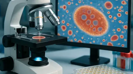
Once collected, the biofluid droplet is placed on a surface and observed as it dries over time, a process captured in real-time using basic brightfield microscopy equipped with a 4x objective lens and a digital camera. This stage is crucial, as the evolving patterns during drying—such as the movement of proteins and cells—reveal intricate details about the sample’s composition. The use of straightforward imaging tools ensures that this method remains practical for widespread application.
Unlocking a Sample’s Hidden Story
Focusing on the dynamic changes during drying, rather than just the final dried pattern, offers a richer understanding of the biofluid’s internal makeup. Each shift in structure as the droplet evaporates tells a unique narrative about the presence of abnormalities or health markers. This real-time observation captures nuances that static analysis might miss, providing a comprehensive diagnostic foundation.
Step 3 – Analyzing Patterns with Machine Learning
The captured time-lapse images of the drying droplet are then processed through advanced machine-learning algorithms, which are trained to identify differences between healthy and abnormal samples. This analytical phase transforms raw visual data into meaningful insights, detecting subtle variations in patterns that indicate specific conditions. The reliance on computational power allows for precision without the need for extensive human interpretation.
Enhancing Accuracy with Artificial Intelligence
Artificial intelligence plays a pivotal role in decoding the complex drying patterns, ensuring a high level of diagnostic accuracy with minimal equipment. By learning from vast datasets of biofluid images, these algorithms can pinpoint irregularities that might escape the untrained eye. This integration of technology enhances reliability, making the system a powerful tool even in settings without access to specialized diagnostic machinery.
Step 4 – Applying the Workflow Across Multiple Biofluids
The final step showcases the system’s versatility, as the same workflow can be applied to analyze various biofluids without requiring adjustments to the process or equipment. Whether testing blood for diabetes markers, saliva for influenza, or urine for malaria indicators, the methodology remains consistent. This adaptability broadens the scope of diseases that can be detected using a single, streamlined approach.
Streamlining Diagnostics for Diverse Applications
The ability to use a universal process across different biofluids translates into significant cost savings and practicality, especially for healthcare providers managing multiple conditions. This unified system reduces the need for distinct testing protocols or additional tools, making it an efficient choice for diverse medical contexts. Such flexibility underscores the technology’s potential to address a wide array of health challenges with ease.
Key Takeaways from the Biofluid Imaging Process
This section condenses the essential elements of the automated biofluid imaging system into a concise overview for quick reference and clarity. Each point highlights a core strength of the technology, demonstrating its value in modern diagnostics.
- Minimal, non-invasive sample collection significantly lowers the burden on patients, fostering greater participation in health checks.
- Real-time imaging of drying droplets uncovers detailed diagnostic insights that static methods cannot match.
- Machine learning delivers precise differentiation between healthy and abnormal samples, ensuring dependable results.
- Versatility across various biofluids enables detection of numerous diseases using the same efficient workflow.
- The use of basic equipment enhances scalability and affordability, making the system viable for global implementation.
Broader Impacts and Future Potential in Global Healthcare
The emergence of automated biofluid imaging aligns seamlessly with the growing demand for equitable healthcare solutions that can operate in diverse settings. By minimizing reliance on costly infrastructure, this technology offers a lifeline to remote and underserved communities where traditional diagnostics are often out of reach. Its potential to deliver laboratory-quality results at the point of care represents a monumental shift in how medical services can be distributed worldwide.
Looking ahead, the system holds promise for further development into portable health-screening tools that could be used directly by individuals or small clinics. Such advancements would empower communities to monitor health proactively, reducing the incidence of late-stage diagnoses. However, challenges remain, including the need to scale production and validate accuracy across varied populations to ensure consistent performance in different environments.
This innovation also ties into a larger trend of integrating technology with healthcare to democratize access to medical care. As machine learning and imaging techniques continue to evolve, the possibility of even more refined and user-friendly diagnostic tools emerges. The ongoing commitment to accessibility and efficiency positions this system as a catalyst for lasting change in global health equity over the coming years, from 2025 onward.
Embracing the Future of Diagnostics with Confidence
Reflecting on the journey of automated biofluid imaging, it becomes evident that each step—from minimal sample collection to machine-learning analysis—works together to simplify and accelerate disease diagnosis. The process not only alleviates the physical and financial burdens of traditional methods but also opens doors to equitable healthcare access. Its adaptability across biofluids and reliance on basic tools marks a turning point in how medical testing is perceived and implemented.
As a next step, stakeholders in healthcare are encouraged to explore partnerships for scaling this technology, ensuring its integration into community health programs worldwide. Investing in further research to refine accuracy and develop portable devices emerges as a critical focus area. Additionally, raising awareness about such innovations proves essential to drive adoption in regions most in need.
Beyond immediate actions, the conversation shifts to envisioning a landscape where diagnostics transcend geographical and economic barriers. The groundwork laid by this system inspires optimism for future tools that could predict and prevent diseases even before symptoms appear. This trajectory of progress highlights a collective responsibility to support and champion accessible medical solutions for all.


