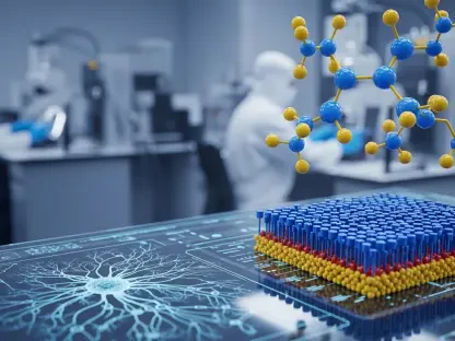

Advancements in cancer therapy have brought significant hope, especially with immune checkpoint blockades (ICBs), providing a groundbreaking treatment strategy for advanced cancers. Despite this, the effectiveness of ICBs has often been thwarted by therapeutic resistance, which renders tumor-infiltrating lymphocytes (TILs) ineffective. Rejuvenating these TILs to restore their anti-cancer abilities has thus become a pivotal goal for oncologists. A critical hurdle in this endeavor is the unique environment within tumors characterized by hypoxia. This low oxygen condition emerges due to the rapid proliferation of cancer cells outpacing the oxygen supply from the often abnormal tumor vasculature. This hypoxic environment profoundly impacts the functioning of T cells, the primary soldiers in our immune defense, within the cancer microenvironment. Delving deep into this issue, researchers have embarked on understanding how hypoxia dictates TIL functionality, aiming to find ways to manipulate these effects and overcome resistance to ICB therapy.
The Role of Hypoxia in Tumor Microenvironment
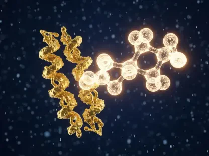
Hypoxia continues to challenge cancer therapy by significantly impacting T cell functions within the tumor microenvironment. Dr. Lewis Zhichang Shi and his team at the University of Alabama at Birmingham have focused on unlocking these intricacies, particularly the role of hypoxia-inducible factors, with a spotlight on HIF1α in dictating T cell activities under hypoxic conditions. HIF1α, a key subunit of the hypoxia-inducible factor (HIF), orchestrates cellular responses to low oxygen levels. Through groundbreaking research, Shi’s team has demonstrated that HIF1α is crucial for the induction of interferon gamma (IFN-γ), a cytokine paramount for the tumor-killing capacity of T cells. Furthermore, the researchers underscored that glycolysis, an alternative metabolic pathway that provides energy in oxygen-deprived conditions, becomes necessary for inducing IFN-γ in T cells amidst hypoxia. This finding highlights a fundamental metabolic reprogramming in T cells under hypoxic stress, a phenomenon pivotal for effective cancer therapies.
Interestingly, under normal oxygen levels (normoxia), HIF1α is not a requisite for IFN-γ induction in T cells. Instead, this function is mediated by LDHa, a downstream target, showcasing a significant metabolic and functional disparity between normoxic and hypoxic conditions. This research bridged a crucial gap by explaining how HIF1α regulation affects both IFN-γ induction and glycolysis in hypoxic T cells. Utilizing advanced genetic mouse models, metabolic flux analysis, including 13C-labeled glucose tracing, and precise pharmacological approaches, the researchers revealed that HIF1α-mediated glycolysis is indispensable for IFN-γ induction under hypoxia. The deletion of the HIF1α gene in both human and mouse T cells halted their metabolic shift from catabolic to anabolic processes, particularly glycolysis, subsequently suppressing IFN-γ production. Pharmacologically inhibiting glycolysis in T cells under hypoxia mirrored this suppression, while stabilization of HIF1α enhanced IFN-γ production, highlighting a critical metabolic control via HIF1α.
Metabolic Shifts in T Cells Under Hypoxia
The impact of hypoxia on the metabolic shifts in T cells introduces a significant dynamic in cancer treatment strategy, further complicating therapeutic approaches. This shift, essentially driven by HIF1α, denotes a critical aspect of T cell metabolism. As opposed to normoxic conditions, where other factors mediate IFN-γ induction, hypoxic environments necessitate HIF1α for this role. Studies detailed how genetic interventions, such as deleting the HIF1α gene, inhibit the metabolic shift vital for T cell activation under low oxygen. Additionally, sophisticated metabolic analyses, including labeling techniques, underscored that HIF1α-driven glycolysis sustains T cell activity in hypoxia, a fundamental insight into metabolic demands during oxygen deprivation.
Further, the pharmacological manipulation of glycolysis corroborated these genetic findings, reinforcing that glycolysis remains essential under hypoxia for proper T cell function. In these studies, knocking out HIF1α or inhibiting glycolysis pharmacologically led to a marked decrease in IFN-γ induction. These findings present a dual-edged sword of metabolic and genetic regulation of T cells in hypoxia. Conversely, enhancing HIF1α by counteracting its negative regulators significantly improved IFN-γ production, underlining strategies to augment T cell functionality amidst hypoxic tumors. This balance of metabolic reprogramming forms a crucial aspect of cancer immunotherapy, emphasizing potential interventions that can restore or boost T cell functions even under challenging hypoxic conditions.
Implications for Cancer Immunotherapy
The profound implications that arise from these studies emphasize the pivotal role of HIF1α in effective cancer immunotherapy. The research highlighted significant challenges and solutions to enhance T cell functionality under hypoxia, illustrating the potential for therapeutic innovations. For instance, hypoxic T cells with deleted HIF1α showcased a diminished capacity to kill tumor cells in vitro while also demonstrating reduced responses to ICB therapy in in vivo models. This starkly contrasted with normal T cells, thus stressing the importance of HIF1α in immune responses within cancer contexts. Revelatory findings by Shi’s team introduced acetate supplementation as a groundbreaking strategy to bypass resistance in these HIF1α-deleted T cells. This supplementation restored glycolytic activity, reigniting IFN-γ production and reinforcing T cell functionality even in hypoxia.
Validation of this strategy in mice further solidified its potential, with acetate supplementation combined with ICB therapy significantly reducing tumor growth and weight in HIF1α-deficient mice. This innovation opens new realms for devising cancer therapies targeting metabolic interventions to boost immune responses. Acetate supplementation exemplifies a tailored approach to reinvigorate T cells, marking a forward stride in cancer therapeutics. The implications extend broadly into clinical practices where these metabolic interventions can be refined and incorporated to enhance patient outcomes, presenting new avenues to overcome existing resistance mechanisms in cancer treatment.
Metabolic Tug-of-War in Tumor Microenvironment
Advancements in cancer treatment have generated significant hope, particularly with the development of immune checkpoint blockades (ICBs), which offer a groundbreaking strategy for treating advanced cancers. However, the success of ICBs is often hindered by therapeutic resistance, rendering tumor-infiltrating lymphocytes (TILs) ineffective. Reinvigorating these TILs to restore their anti-cancer functions has become a critical goal for oncologists. One major obstacle in this effort is the unique tumor environment, particularly its hypoxic (low oxygen) conditions. This hypoxia arises because the rapid growth of cancer cells surpasses the oxygen supply from the often irregular tumor blood vessels. This low oxygen condition heavily affects the performance of T cells, the main components of our immune defense within the tumor’s microenvironment. Researchers are delving into how hypoxia controls TIL functionality, aiming to discover ways to counteract these effects and defeat resistance to ICB therapy. Their findings could pave the way for enhancing the efficacy of cancer immunotherapy.