
Ivan Kairatov is a leading biopharma expert with a distinguished background in structural biology and drug development, currently focusing on the mechanics of cellular signaling. His work bridges the gap between high-resolution molecular imaging and the practical application of these insights to treat human disease. In this discussion, we explore the groundbreaking visualization of the iRhom1-ADAM17 complex, a discovery that clarifies how cells communicate during inflammatory responses and highlights why human biology sometimes deviates significantly from long-standing animal models.
The conversation covers the mechanics of cryogenic electron microscopy in identifying molecular relays, the functional divergence between nearly identical proteins, and the therapeutic potential of targeting regulator proteins to mitigate chronic inflammation.
Cryogenic electron microscopy allows for high-resolution visualization of protein complexes like iRhom1 and ADAM17. How did this technology help identify the structural elements of the molecular relay, and what specific steps were involved in capturing these images to reveal the link between intracellular signaling and surface activation?
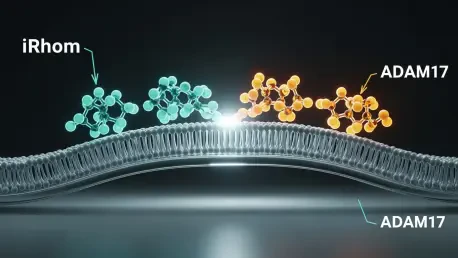
The use of cryogenic electron microscopy, or cryo-EM, was absolutely transformative for our ability to see these proteins in their native-like states without the distortions common in older methods. By flash-freezing the iRhom1-ADAM17 complex, we were able to capture high-resolution snapshots that revealed the specific structural elements acting as a molecular relay across the cell membrane. This process involved purifying the complex and stabilizing it in a thin layer of vitreous ice, allowing us to observe how signals from inside the cell are physically transmitted to the surface-level enzymes. Seeing these structures for the first time allowed us to map the precise path that links intracellular signaling networks to the activation of ADAM17. It is a level of detail that has eluded the scientific community for 30 years, finally showing us the “bridge” that allows these two components to talk to one another.
Ectodomain shedding allows enzymes like ADAM17 to cleave surface proteins and alter cell communication. How does the iRhom1-ADAM17 complex facilitate this process during tissue development, and what metrics or benchmarks indicate whether this activity is functioning within a healthy range for the human immune response?
The iRhom1-ADAM17 complex acts as the primary driver for ectodomain shedding, a process where the enzyme acts like a pair of molecular scissors to release protein targets from the cell surface. This shedding is vital because it triggers rapid changes in cell-to-cell communication, which is a fundamental requirement for healthy tissue development and a robust immune response. When the system is functioning correctly, we see a balanced release of these signaling proteins; however, if the activity becomes dysregulated, it can lead to chronic inflammation or even cancer. The healthy range is maintained by the iRhom proteins acting as “master regulators,” ensuring ADAM17 only cleaves its targets when the proper intracellular signals are received. We look for the successful transport of ADAM17 to its residence at the cell surface as a key benchmark for healthy cellular function.
Both iRhom1 and iRhom2 share identical structures and signal responses, yet they perform divergent roles in substrate recognition. How do these proteins differentiate their tasks despite these similarities, and what sequence nuances or environmental factors allow them to make these distinct functional decisions at the cell surface?
It is a fascinating paradox in structural biology that iRhom1 and iRhom2 appear identical in their overall architecture and respond to the same signals, yet they choose different substrates to cleave. We believe the secret lies in the subtle nuances of their amino acid sequences, which act as a fine-tuned recognition system for specific protein targets. While the “body” of the protein looks the same, these small sequence variations allow each iRhom to preferentially recruit and activate ADAM17 for different tasks. This allows the cell to use a unified model for enzyme activation while still maintaining highly specific control over which messages are sent to neighboring cells. Understanding why they make these different decisions is the next great frontier in our research, as it represents the missing piece of the puzzle in how inflammatory pathways are governed.
Mutations in iRhom1 are linked to conditions like cardiomyopathy where proteins fail to fold properly. What happens to the ADAM17 enzyme when its regulator cannot reach the cell surface, and how do these human outcomes differ from the phenotypes traditionally observed in previous animal models?
When a mutation occurs, such as those seen in patients with cardiomyopathy, the iRhom1 protein fails to fold into its correct three-dimensional shape, rendering it functionally null. Because ADAM17 exists only as a complex with these iRhom proteins, the enzyme is effectively trapped inside the cell and can never reach its target area near the surface. This total loss of function means the essential shedding process never happens, leading to severe developmental and cardiac issues. Interestingly, the human outcomes we are seeing are quite distinct from what was previously documented in animal studies. This suggests that human iRhom1 biology has unique complexities, and these new structural insights give us the first real understanding of how these disease phenotypes manifest specifically in our own species.
Targeting iRhom proteins is a potential strategy for treating chronic inflammatory diseases and neurodegenerative disorders. What is the step-by-step process for leveraging iRhom2 specificity to regulate ADAM17, and how might targeting the regulator rather than the enzyme itself reduce the risks of dysregulated activity?
The strategy involves shifting our focus away from the ADAM17 enzyme itself—which is difficult to target without causing broad side effects—and instead focusing on the iRhom2 regulator. By developing drugs that specifically bind to the iRhom2-ADAM17 complex, we can selectively dampen the inflammatory signals driven by that specific pairing while leaving other essential functions of ADAM17 intact. This step-by-step approach starts with using our high-resolution cryo-EM maps to identify unique binding pockets on iRhom2 that are absent in other proteins. Targeting the regulator provides a much more surgical level of control, reducing the risk of the widespread, “off-target” dysregulated activity that often plagues traditional enzyme inhibitors. It is a more sophisticated way to dial down inflammation without compromising the patient’s overall immune health.
What is your forecast for the treatment of chronic inflammatory diseases?
I believe we are entering an era of “regulator-centric” therapy where we will no longer attempt to shut down entire enzymes, but rather modulate the specific proteins that tell them when to work. With our new ability to visualize the iRhom1 and iRhom2 complexes at an atomic level, I forecast the development of a new generation of highly specific inhibitors that can treat conditions like rheumatoid arthritis or neurodegeneration with far fewer side effects. We are moving away from the “blunt instrument” approach of the last 30 years and toward a future where we can precisely tune the molecular relays of the cell. Within the next decade, I expect these structural insights to yield clinical candidates that finally address the root cause of dysregulated shedding in human patients.