
The delicate balance between eradicating a life-threatening malignancy and preserving the fundamental structural integrity of the human heart has long remained one of the most perilous tightropes in modern medicine. While the introduction of immune checkpoint inhibitors (ICIs) has fundamentally altered the survival landscape for patients with advanced cancers, this revolutionary therapy often acts as a double-edged sword. The same mechanisms that empower the immune system to dismantle a tumor can, without warning, provoke the body’s defenses to turn against its own cardiovascular system.
For years, the clinical community has grappled with the devastating trade-offs inherent in modern immunotherapy. Patients who successfully fought off aggressive malignancies were frequently met with a second, equally dangerous foe: heart failure induced by treatment-related inflammation. The absence of a sensitive, real-time monitoring tool meant that these side effects often remained invisible until irreversible damage had already occurred. However, a transformative shift in molecular imaging is now providing a way to watch both the cancer’s retreat and the heart’s distress through a single, comprehensive lens.
By focusing on a specific molecular signature that links oncological activity to cardiovascular decay, researchers have unlocked a method to predict and prevent these complications before they become life-threatening. This advancement does more than just diagnose; it creates a dynamic dashboard for personalized medicine. It allows physicians to fine-tune therapies in real-time, ensuring that the pursuit of a cancer-free life does not come at the cost of a failing heart.
The High Stakes of the Modern Medical Paradox
Immunotherapy has rewritten the rules of cancer survival, yet this medical triumph carries a heavy price for the human heart. While immune checkpoint inhibitors are remarkably effective at mobilizing the body’s defenses to dismantle tumors, they frequently trigger unintended inflammatory attacks on the cardiovascular system. This phenomenon presents a gut-wrenching choice for clinicians and patients alike: continue a life-saving cancer treatment at the risk of permanent heart damage, or halt therapy and allow the malignancy to regain its foothold.
This biological paradox stems from the way these drugs interact with the immune system’s regulatory checkpoints. By removing the “brakes” that prevent T-cells from attacking the body, ICIs allow for a robust assault on cancer cells that have previously evaded detection. Unfortunately, the heart and vascular tissues often become collateral damage in this heightened immune state. The resulting inflammation can lead to acute crises, such as myocarditis, or long-term chronic issues like the rapid progression of arterial disease.
The stakes are particularly high for patients who already possess underlying cardiovascular risk factors. For these individuals, the immune response is not just a localized battle against a tumor; it is a systemic event that can destabilize existing atherosclerotic plaques or trigger inflammation in the heart muscle. Consequently, the medical community has urgently sought a way to bridge the gap between effective oncology and cardiovascular safety.
The Rise of Cardio-Oncology and the Threat of Immune-Related Side Effects
The emergence of cardio-oncology as a specialized field highlights a growing concern regarding immune-related adverse events. For a significant number of patients, the “brakes” removed from the immune system stay off, leading to myocarditis, accelerated plaque buildup in the arteries, and a heightened risk of heart attacks. Until recently, the medical community lacked the tools to catch these subclinical changes early enough to intervene. Without a sensitive, noninvasive way to monitor both the cancer’s retreat and the heart’s distress simultaneously, doctors have been forced into a reactive mode.
Traditional monitoring techniques, such as echocardiograms or blood tests for troponin levels, often only reveal damage after it has reached a critical threshold. These methods lack the molecular specificity required to distinguish between general stress and the specific inflammatory pathways activated by immunotherapy. Because cardiovascular side effects can manifest silently, the lack of early warning systems often results in patients presenting with advanced heart failure symptoms long after the window for preventative intervention has closed.
The challenge is compounded by the fact that many of the symptoms of cardiac distress, such as fatigue or shortness of breath, can be easily mistaken for general side effects of cancer or the cancer treatment itself. This diagnostic ambiguity places a massive burden on cardio-oncologists to determine whether a patient’s heart is genuinely under threat. To move forward, the field required a breakthrough that could visualize the actual movement of inflammatory cells within the vascular system.
Visualizing Progress with CCR2: A Dual Beacon for Cancer and Heart Health
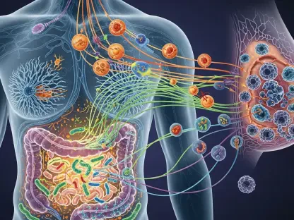
The breakthrough lies in a specific molecular target known as the C-C chemokine receptor type 2 (CCR2). This protein acts as a common denominator between oncological progression and cardiovascular decay, as it is expressed in both the tumor microenvironment and the inflammatory cells within atherosclerotic plaques. By utilizing the novel radiotracer 64Cu-DOTA-ECL1i, researchers can now perform a single PET scan that serves two purposes. A high signal within the tumor indicates a robust immune response against the cancer, while a rising signal in the heart or aorta acts as an early warning system for dangerous inflammation.
The CCR2 receptor is a primary driver of the recruitment of monocytes and macrophages, which are the specialized cells that fuel inflammation in both cancer sites and damaged vascular tissues. When the 64Cu-DOTA-ECL1i tracer is injected into a patient, it binds specifically to these CCR2-expressing cells. This binding creates a high-contrast image on a PET scan, allowing clinicians to see exactly where the immune system is most active. It effectively turns the patient’s own immune response into a visible map that doctors can follow throughout the course of treatment.
This dual-utility transforms the PET scanner into a comprehensive dashboard for patient health. Instead of ordering multiple disparate tests, a single imaging session provides data on how the tumor is responding to treatment while simultaneously assessing the status of the patient’s arteries and heart muscle. This holistic view is essential for navigating the complex interactions between the immune system and the various organ systems affected by systemic therapy.
Evidence from the Lab: Quantifying Cardiac Risks and Tracer Efficacy
Research led by Dr. Jaume Otaegui at Washington University provided clear, quantifiable evidence of this technology’s potential. In comparative studies, the CCR2-targeted tracer significantly outperformed the traditional imaging standard, 18F-FDG, providing much sharper visualizations of both tumors and vascular inflammation. The study’s most striking finding revealed that while standard immunotherapy successfully shrunk tumors, it also caused a measurable spike in cardiac inflammation—a “cardiac cost” that was previously difficult to see in real-time.
To achieve these results, the research team utilized a sophisticated mouse model that replicated the conditions of an older adult patient who might have both cancer and underlying cardiovascular disease. They observed that when the mice were treated with standard checkpoint inhibitors, the PET scans showed a distinct and rising signal in the heart and the aorta. This was not a random occurrence; the imaging proved that the treatment was actively recruiting inflammatory cells into the heart’s sensitive tissues even as it successfully inhibited tumor growth.
Notably, the study demonstrated that the 64Cu-DOTA-ECL1i tracer had a much higher affinity for its target cells than the glucose-based tracers used in standard PET scans. While 18F-FDG is often obscured by the heart’s natural metabolic activity, the CCR2-targeted tracer remained clear, providing a precise measurement of the actual inflammatory load. This research confirmed that the inflammatory response isn’t just a side effect; it is a measurable physiological event that can be anticipated and tracked with scientific precision.
A Proactive Strategy for Balancing Immunotherapy and Cardiovascular Protection
Moving beyond diagnosis, the latest findings suggested a practical framework for mitigating these risks through synergistic therapy. By combining immunotherapy with itacitinib—a JAK1 inhibitor—clinicians were able to shield the heart without compromising cancer treatment. In experimental models, this combination not only reduced the inflammatory CCR2 signal in the heart and aorta but actually enhanced the body’s ability to control the tumor. This strategy offered a clear path forward: use CCR2 PET imaging to monitor the heart’s status throughout treatment and introduce protective agents like itacitinib the moment inflammation is detected.
The discovery that a JAK1 inhibitor could actually improve the tumor-fighting capability of the immune system while protecting the heart was a significant revelation for the field of cardio-oncology. It suggested that inflammation is not a monolithic force; rather, it can be modulated to favor the destruction of cancer while sparing healthy tissues. This nuanced approach allowed for the continuation of aggressive cancer treatments that would have otherwise been deemed too risky for the patient’s cardiovascular health.
Ultimately, the shift from reactive to proactive care ensured that the pursuit of a cancer cure no longer required the sacrifice of cardiovascular health. The study established that by integrating molecular imaging with targeted anti-inflammatory drugs, the medical community could provide a safer, more effective route to survival. This integrated model of care represented a landmark achievement in precision medicine, offering patients a way to fight cancer without compromising the very organ that sustains their life. This transition toward sophisticated, real-time monitoring signaled a new era where the heavy price of survival was finally being reduced.