
The traditional view of acute myeloid leukemia as a localized disease of the bone marrow is undergoing a radical transformation as researchers uncover the sophisticated ways this cancer colonizes distant organs like the lungs. While clinical experts have long recognized that respiratory failure is a primary cause of early mortality in patients with this blood cancer, the precise biological mechanisms driving this organ-specific destruction remained largely mysterious until very recently. This article aims to explore the groundbreaking findings that illustrate how leukemia cells act as biological architects, actively remodeling the lung microenvironment to favor their own survival at the expense of the patient’s ability to breathe. Readers can expect to learn about the specific cellular transformations, the inflammatory pathways involved, and the emerging therapeutic targets that may soon change the standard of care for these high-risk clinical scenarios.
The scope of this discussion encompasses the transition of leukemia from a marrow-centric disease to a systemic one that specifically compromises the structural and immune integrity of pulmonary tissue. By examining the results of high-resolution multi-omic studies, we can see how malignant cells do not just inhabit the lungs but systematically dismantle the very cells responsible for gas exchange. This analysis provides essential context for understanding why traditional leukemia treatments sometimes fail to address respiratory distress and how new strategies, including the use of common corticosteroids and experimental antibodies, offer a path toward better patient outcomes. Through this exploration, the complex dialogue between the tumor and its surrounding environment becomes a map for future medical intervention.
Key Questions or Key Topics Section
What Factors Make the Lung a Prime Target for Leukemic Migration?
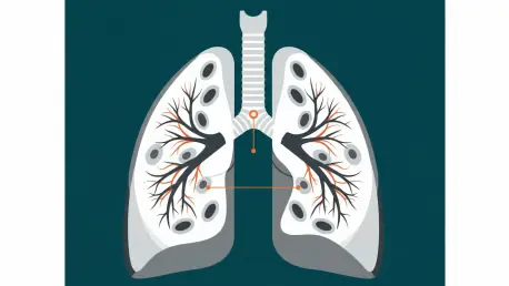
The migration of acute myeloid leukemia cells from the bone marrow into the peripheral organs is not a random occurrence but a calculated movement facilitated by the body’s vascular network. Researchers have discovered that leukemia cells possess a specific affinity for the lungs, utilizing the bloodstream as a high-speed corridor to reach pulmonary tissues. This preference is particularly dangerous because the lungs are highly vascularized, providing a vast surface area for malignant cells to anchor and begin their colonization process. Understanding this migratory pattern is vital for clinicians because it explains why respiratory symptoms often appear early in the disease progression, sometimes even before a formal diagnosis is established in the bone marrow.
Once these malignant cells arrive in the pulmonary environment, they gravitate toward the alveolar and perivascular regions, which are the most critical zones for oxygenation. By employing advanced technologies like single-cell and spatial transcriptomics, scientists have been able to observe this infiltration with unprecedented clarity. The leukemia cells do not simply sit in the vessels; they actively cross the vascular barriers to enter the lung stroma. This movement creates a localized “niche” where the cancer can hide from systemic treatments while simultaneously beginning the process of tissue degradation that leads to the clinical presentation of respiratory failure.
The importance of this specific organ targeting lies in the unique vulnerability of the lung’s architecture compared to other soft tissues. Unlike the liver or spleen, which can often tolerate a degree of infiltration without immediate functional collapse, the lungs rely on incredibly thin membranes for gas exchange. When leukemia cells concentrate in these areas, they disrupt the delicate balance required for breathing. This insight has shifted the research focus toward identifying the specific molecular “homing” signals that attract these cells to the lungs, with the goal of developing therapies that can block this migration before the organ’s structural integrity is compromised.
How Does Leukemia Physically Dismantle the Architecture of the Lungs?
The presence of leukemia cells in the lungs initiates a destructive process that goes far beyond simple overcrowding. As these malignant cells infiltrate the tissue, they cause significant damage to the capillaries and the epithelial lining of the alveoli. This damage increases vascular permeability, leading to a condition where fluid leaks into the air sacs, making it difficult for the patient to inhale effectively. More alarmingly, the study of this phenomenon has revealed a rapid depletion of specialized cells known as capillary aerocytes and type 1 alveolar epithelial cells. These are the primary structural components responsible for the efficient transfer of oxygen into the blood and carbon dioxide out of it.
Beyond the immediate loss of functional cells, the leukemia-driven environment triggers a pathological repair response that results in permanent scarring. The research identified an abnormal increase in fibroblasts and myofibroblasts, which are cells that typically help heal wounds. However, under the influence of the leukemic microenvironment, these cells begin to overproduce collagen, leading to extensive fibrotic remodeling. This stiffening of the lung tissue means the organ loses its elasticity, forcing the body to work significantly harder to move air. Functional tests in experimental models confirmed that this structural stiffness directly correlates with the severity of respiratory distress seen in human patients.
This architectural collapse represents a dual threat: the loss of the cells that perform gas exchange and the physical hardening of the organ itself. The transition from a flexible, air-filled structure to a dense, scarred environment happens with surprising speed during the early stages of leukemic infiltration. By recognizing that the damage is both cellular and structural, the medical community is moving toward a more holistic view of leukemia management. Addressing the cancer alone is no longer seen as sufficient; preserving the physical architecture of the lungs has become a parallel priority in preventing the fatal complications associated with the disease.
Why Does the Inflammation Caused by Leukemia Mimic Severe Viral Infections?
One of the most striking discoveries in recent pulmonary research is that the inflammation triggered by acute myeloid leukemia is not a localized reaction but an organ-wide crisis. The pattern of this inflammation bears an uncanny resemblance to the “cytokine storm” observed in patients with severe respiratory viruses, such as COVID-19. In both cases, the lungs become flooded with inflammatory proteins like Tumor Necrosis Factor and various interleukins. This widespread immune activation creates a toxic environment that damages healthy tissue throughout the lung, regardless of whether leukemia cells are physically present in every single area of the organ.
This similarity to viral infections is important because it highlights a shift in how we understand cancer-related inflammation. In traditional solid tumors, the inflammatory response is usually localized to the immediate vicinity of the mass. In contrast, leukemia creates a systemic and pervasive inflammatory state within the lung that leads to the depletion of vital immune “sentinel” cells, such as T cells and Natural Killer cells. This depletion leaves the lung vulnerable and unable to mount a specific defense against the cancer. The presence of high levels of inflammatory monocytes further exacerbates the tissue damage, mirroring the pathological pathways seen in acute respiratory distress syndrome.
The implications of this viral-like inflammation are profound for clinical diagnosis and treatment. Patients presenting with sudden respiratory failure and high levels of inflammatory markers might be misdiagnosed as having a primary infection if the underlying leukemia is not considered. Furthermore, the identification of this cytokine storm provides a scientific basis for using anti-inflammatory drugs that are typically reserved for viral crises. By dampening this excessive immune response, doctors may be able to stabilize the patient’s respiratory function long enough for chemotherapy to begin its work on the primary cancer.
How Does the Disease Subvert the Local Immune Response Within Lung Tissue?
The leukemic microenvironment is not just a site of destruction; it is a masterclass in immune evasion and subversion. Leukemia cells effectively “re-tune” the lung’s immune system to work in their favor. While the lungs are normally guarded by a balanced population of lymphocytes and macrophages that protect against pathogens and abnormal cells, the infiltration of leukemia skews this population toward a pro-inflammatory but anti-tumor state. There is a massive surge in activated neutrophils and nonclassical monocytes which, rather than attacking the cancer, contribute to the hyper-inflammatory environment that facilitates further tumor expansion and tissue damage.
At the same time that these inflammatory cells are increasing, the adaptive immune system is being systematically silenced. The cells responsible for recognizing and killing malignant cells—T cells and B cells—are significantly reduced in number and function. This creates a “double-blind” scenario where the lung is simultaneously overwhelmed by non-specific, damaging inflammation and paralyzed when it comes to the specific immune response needed to clear the leukemia. This immune reorganization ensures that the leukemia cells can continue to proliferate without being checked by the body’s natural defense mechanisms, essentially turning the lung into a sanctuary for the disease.
Moreover, the communication between the leukemia cells and the remaining lung cells is highjacked. Malignant cells emit signals that keep the local macrophages in a state that supports tumor growth rather than tumor suppression. This sophisticated level of control over the immune landscape is why leukemia can be so difficult to treat once it has established itself in the lungs. By understanding these specific immune shifts, researchers are now looking for ways to “re-set” the pulmonary immune system, hoping to restore the natural balance and allow the body’s own defenses to assist in the fight against the cancer.
Which Molecular Toolkits Represent the Most Promising Therapeutic Targets?
Mapping the communication pathways between leukemia and the lung has revealed two specific molecular targets that seem to drive the disease’s success in the pulmonary environment. The first is a protein called Galectin-9, which is encoded by the LGALS9 gene. This protein is highly expressed in leukemia cells and acts as a bridge, allowing the tumor to interact with and suppress the lung’s immune cells. In clinical studies, patients with high levels of this protein consistently showed worse survival rates and more severe lung inflammation, making it a prime candidate for targeted antibody therapy designed to break the leukemia’s hold on the tissue.
The second critical pathway involves a “danger signal” cytokine known as IL-33 and its receptor, IL1RL1. When the structural cells of the lung are damaged by infiltrating leukemia, they release IL-33 as a distress signal. In a cruel twist, the leukemia cells express the receptor for this signal and use it as a growth factor to fuel their own expansion. This feedback loop—where the damage caused by the cancer actually provides the fuel for more cancer—is a hallmark of the disease’s predatory nature. Blocking this receptor in experimental settings has shown that the cycle can be broken, leading to a significant reduction in the tumor burden and an increase in leukemia cell death.
These discoveries are exciting because they offer “vulnerable points” in a disease that has historically been very difficult to manage. By targeting Galectin-9 or the IL-33 axis, physicians may soon be able to use combination therapies that specifically protect the lungs while the leukemia is being treated systemically. This would represent a shift from broad-spectrum chemotherapy to a more nuanced approach that addresses the specific ways the cancer interacts with different organs. As these targets move toward clinical trials, they provide hope for a future where respiratory failure is no longer a guaranteed death sentence for AML patients.
What Role Does Corticosteroid Treatment Play in Managing Respiratory Failure?
The use of glucocorticoids like prednisone has emerged as a surprisingly effective tool in the immediate management of leukemia-induced respiratory failure. While some clinicians have used steroids empirically in the past, new research provides the scientific rationale for why they work so well. Prednisone acts by rapidly dampening the “inflammatory fire” in the lungs, reducing the accumulation of leukemia cells in the capillaries and the surrounding spaces. By cooling the cytokine storm, the drug allows the surviving lung structures to function more efficiently almost immediately, providing a critical window for more definitive cancer treatments to be administered.
Retrospective data from human patients has shown that the response to prednisone can be remarkably fast. In several documented cases, patients with severe respiratory distress showed improvements in their oxygen requirements within just 12 hours of receiving the medication. By the 24-hour mark, many patients had seen their supplemental oxygen needs drop by as much as 75%. This rapid physiological improvement is not just about symptom management; it actually changes the physical landscape of the lung, reducing the density of malignant cells and clearing the way for better gas exchange.
However, the medical community also emphasizes the need for caution and precise diagnosis before deploying this strategy. Because corticosteroids suppress the immune system, they could be dangerous if the patient’s respiratory failure is caused by an underlying infection rather than the leukemia itself. Therefore, the successful use of prednisone in this context requires sophisticated diagnostic tools to ensure that the lung distress is truly a result of leukemic infiltration. When used correctly, this approach represents a bridge to survival, stabilizing the most vulnerable patients during the most dangerous phase of their illness.
Summary or Recap
The transformation of the lung microenvironment by acute myeloid leukemia is a multifaceted process involving physical infiltration, structural destruction, and immune subversion. We have seen how leukemia cells migrate toward the lungs and dismantle the aerocytes and epithelial cells necessary for breathing, while simultaneously inducing fibrosis that scars the tissue. The resulting environment mimics the hyper-inflammation of a viral crisis, where the immune system is “re-tuned” to protect the cancer rather than destroy it. Key molecular targets like Galectin-9 and the IL-33 receptor offer new hope for targeted therapies that can break the pathological dialogue between the tumor and the organ. Furthermore, the strategic use of prednisone has been validated as a life-saving intervention for managing the acute respiratory complications of the disease. These insights underscore the importance of viewing leukemia as a systemic architect of disease, requiring a multi-faceted treatment strategy that addresses both the malignant cells and the organs they inhabit.
Conclusion or Final Thoughts
The scientific community previously regarded the lungs as passive victims of leukemic spread, but the evidence gathered in recent years established them as active, though unwilling, participants in the disease’s progression. Researchers observed that the interaction between malignant blasts and pulmonary tissue created a unique pro-tumor niche that demanded specialized intervention strategies beyond traditional bone marrow treatments. This paradigm shift suggested that future protocols must prioritize organ-specific protection to mitigate the high mortality rates associated with respiratory failure. As medical professionals looked toward the next generation of therapies, the focus moved toward clinical trials that combined systemic chemotherapy with localized immune modulators and anti-fibrotic agents. The study of these intricate biological relationships provided a roadmap for turning a devastating complication into a manageable aspect of the disease, ultimately aiming to ensure that no patient lost their life simply because their lungs could no longer bridge the gap between oxygen and the blood.