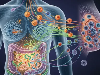

The relentless pursuit of a cancer treatment that spares the patient while obliterating the tumor has led researchers to an architectural marvel smaller than a speck of dust. Modern oncology faces a persistent paradox where the very drugs designed to save lives often cause profound systemic damage due to their non-selective nature. While traditional chemotherapy remains a primary weapon against malignancy, its inability to distinguish between a tumor and healthy tissue frequently leads to debilitating side effects that ravage the human body. This “scorched earth” approach limits treatment dosages and compromises patient well-being, driving researchers at the Indian Institute of Technology Gandhinagar (IITGN) to seek a more surgical alternative.
By pivoting toward the field of DNA nanotechnology, scientists are now exploring ways to deliver therapeutic payloads with unprecedented precision, potentially ending the era of collateral damage in cancer treatment. The current strategy focuses on minimizing the contact between potent drugs and healthy somatic cells, thereby reducing the systemic toxicity that defines the patient experience in traditional wards. This shift represents a move from broad-spectrum chemical warfare toward a refined molecular strategy that prioritizes the integrity of the host while ensuring the total destruction of the target.
Breaking the “Scorched Earth” Cycle: Traditional Chemotherapy
The fundamental challenge in cancer treatment is the biological similarity between malignant cells and the healthy ones that surround them. Traditional chemotherapeutic agents are designed to target rapidly dividing cells, a characteristic of cancer, but one that is also shared by hair follicles, bone marrow, and the lining of the digestive tract. Consequently, patients often experience a cascade of secondary health issues, ranging from immune suppression to organ failure, which can be as life-threatening as the primary diagnosis. This lack of specificity necessitates a delivery system that can navigate the bloodstream without discharging its cargo prematurely.
The high toxicity of these drugs often forces clinicians to walk a tightrope, balancing a dose high enough to kill the cancer but low enough to keep the patient alive. This narrow therapeutic window frequently results in sub-optimal treatment outcomes or the development of drug-resistant tumors. By developing nanostructures that shield the drug until it reaches its destination, the researchers at IITGN aim to widen this window, allowing for more aggressive treatment of the tumor with significantly fewer side effects. The goal is to transform cancer from a systemic trauma into a localized, manageable condition.
Transforming Genetic Blueprints: Programmable 3D Nanostructures
DNA is widely recognized as the fundamental code of life, but in the realm of nanomedicine, it serves as a remarkably versatile and programmable engineering material. Through the principles of self-assembly, scientists can fold specific DNA sequences into complex, three-dimensional shapes that act as microscopic delivery vehicles. The DNA tetrahedron, a stable and pyramid-like structure, has emerged as a frontrunner because its geometry provides exceptional structural integrity while remaining entirely biocompatible within the human body. These scaffolds are inherently less likely to trigger immune rejection compared to synthetic alternatives, making them ideal candidates for the next generation of medical interventions.
Despite these advantages, the primary obstacle in the development of these nanostructures has always been cellular entry. The protective lipid membranes of human cells are designed to keep foreign materials out, and DNA tetrahedrons often struggle to bypass this natural barrier on their own. This hurdle has historically limited the effectiveness of DNA-based delivery systems in clinical settings. However, the programmable nature of DNA allows for the attachment of various functional groups that can modify the physical and chemical properties of the tetrahedron, effectively “tuning” it for better performance in biological environments.
The Synergistic Power: Alpha-Tocopherol and DNA Nanotechnology
To overcome the persistent barrier of cellular internalization, the team at IITGN functionalized these DNA tetrahedrons with alpha-tocopherol succinate (αT), a specialized derivative of Vitamin E. This modification transforms the delivery vehicle from a simple passive carrier into an active and intelligent therapeutic participant. The αT molecule acts as a lipophilic “key,” increasing the structure’s affinity for the fatty cell membrane and facilitating easier entry into the cancerous target. This clever use of biochemistry allows the nanostructure to “melt” through the cell’s outer defenses, ensuring that the therapeutic payload is delivered directly to the intracellular environment.
Beyond acting as a guide, this vitamin derivative possesses a unique biological profile that allows it to selectively disrupt metabolic functions in cancer cells while providing antioxidant protection to healthy ones. This creates a dual-action system where the delivery mechanism itself contributes to the destruction of the malignancy. The synergy between the DNA scaffold and the αT modification ensures that the treatment is not only more efficient but also inherently safer for the surrounding tissue. This approach moves away from the idea of a “dumb” carrier toward an integrated system where every component serves a biological purpose.
Measuring Success: Selective Cytotoxicity and Oxidative Stress
Rigorous testing using methods such as Dynamic Light Scattering (DLS) and fluorescence imaging has confirmed that these αT-modified tetrahedrons significantly outperform standard nanostructures. DLS allowed the researchers to measure the hydrodynamic size and stability of the particles, ensuring they remain intact throughout their journey in the body. Once these structures penetrate the cancer cell membrane, they trigger a lethal biological cascade that specifically targets the metabolic vulnerabilities of malignant cells. The specificity of this interaction is the cornerstone of the research, as it demonstrates a clear preference for diseased tissue over healthy control groups.
Inside the cancerous environment, these nanostructures stimulate the overproduction of Reactive Oxygen Species (ROS), leading to an extreme state of oxidative stress that shatters the cancer cell’s DNA and compromises its mitochondria. This process culminates in apoptosis, a form of programmed cell suicide, ensuring the cancer cell is neutralized without leaking harmful contents into the surrounding healthy tissue. Lead author P Chithra and Dr. Raghu Solanki noted that these findings validated how subtle surface-level modifications could fundamentally rewrite the interaction between nanomaterials and biological systems. This selective cytotoxicity is what differentiates this technology from the indiscriminate nature of traditional chemotherapy.
A Strategic Roadmap: Future Nanomedicine Development
The transition of this breakthrough from a controlled laboratory setting toward a hospital bedside required a structured and meticulous developmental framework. The path forward involved moving beyond in vitro experiments to evaluate how these nanostructures navigated the complex environments of living organisms through advanced animal models. Key steps established comprehensive safety profiles to ensure long-term degradation remained non-toxic while initiating the first phases of clinical trials to determine precise human dosages. This roadmap provided a blueprint for the rational design of future therapies where molecular engineering allowed for the navigation of the human body with the intent of eliminating disease with surgical accuracy.
The scientific community recognized that the success of these DNA-based platforms depended on scalable manufacturing and consistent quality control. Researchers focused on refining the self-assembly process to produce uniform tetrahedrons in large quantities, a necessity for widespread clinical adoption. Furthermore, the integration of real-time tracking modules within the DNA structures allowed for the monitoring of drug release and distribution in vivo. These advancements transformed the initial laboratory discovery into a robust therapeutic candidate that offered hope for a more compassionate era of oncology. Through these efforts, the foundation was laid for a new standard of care where the treatment became as precise as the diagnosis itself.