
Artificial intelligence (AI) has garnered significant attention in recent years, particularly within the medical field for its potential to revolutionize various aspects of healthcare. The integration of AI in breast cancer detection promises not only an increase in accuracy but also a potential reduction in the extensive workload radiologists face. This article delves into a comprehensive study that analyzed the effects of AI integration in mammography screening, highlighting its impact on breast cancer detection rates and radiologist workload within Germany’s nationwide breast cancer screening program.
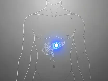
Mammography screening, a vital method for early breast cancer detection, significantly reduces breast cancer-related mortality rates. This process involves the detailed interpretation of mammograms by radiologists, who must review a large volume of images regularly. The repetitive nature of this task, combined with the necessity for high sensitivity and specificity, places a substantial burden on radiologists. This workload is likely to increase further with expanded mammography guidelines that could include broader age groups. Thus, the potential of AI to alleviate this burden, increasing efficiency and accuracy, is particularly compelling.
The Role of AI in Mammography Screening
Enhancing Cancer Detection Rates
Recent studies have shown that AI can match or even exceed the accuracy of radiologists in detecting breast cancer, which can lead to higher cancer detection rates and a reduction in unnecessary patient recalls. However, previous studies had limitations, including small sample sizes and inconsistencies among radiologists, screening sites, and equipment vendors, affecting the generalizability of their findings. To address these limitations, a large-scale, real-world study was conducted to implement AI in a population-based mammography screening program.
This extensive study included data from 461,818 women aged 50-69, who participated in Germany’s nationwide breast cancer screening program between July 2021 and February 2023. All participants underwent four mammograms, which were initially interpreted independently by two radiologists. When either radiologist suspected a case of cancer, a consensus conference was held to decide if further assessments were necessary. This process aimed to maintain high diagnostic accuracy while recognizing the additional workload it created for radiologists.
AI Technology and Its Features
In the screening program, AI technology was utilized to support and enhance the process of mammogram evaluation. The participants were divided into two groups: the AI group and the control group. The AI group employed the AI-supported viewer, Vara MG, which incorporated two primary features: normal triaging and a safety net. Normal triaging was designed to automatically classify highly unsuspicious exams as normal, effectively reducing the number of images radiologists had to review in detail.
The safety net function of Vara MG provided another layer of security by highlighting highly suspicious cases and assisting in localizing potentially cancerous regions on the mammogram. This feature prompted radiologists to give additional attention to flagged exams, thereby potentially increasing the accuracy of cancer detection. By integrating these features, the AI system aimed to streamline the workflow for radiologists while maintaining or improving diagnostic precision.
Impact on Radiologist Workload
Workload Reduction
The results of the study demonstrated a notable impact of AI integration on the workload of radiologists. With AI classifying 59.4% of mammograms as normal, there was a significant potential reduction of about 43% in the time radiologists spent interpreting normal cases. This allowed radiologists to devote more time and attention to complex analyses, possibly leading to an overall increase in diagnostic precision and improved patient outcomes.
The breast cancer detection rate (BCDR) in the AI-supported group was higher compared to the control group, with the AI group achieving a detection rate of 6.7 per 1,000 women as opposed to 5.7 per 1,000 women in the control group. This increase in detection rates suggests that AI can aid radiologists in identifying more instances of breast cancer than traditional methods alone.
Improved Efficiency
One of the most significant findings of the study was the substantial increase in the detection of ductal carcinoma in situ (DCIS) within the AI group. The detection rates rose from 0.8 per 1,000 women in the control group to 1.4 per 1,000 women in the AI group. While the earlier detection of DCIS is promising, it also brought concerns regarding possible overdiagnosis and overtreatment. Not all cases of DCIS progress to invasive cancer, and this aspect will require careful consideration in future AI system implementations.
In terms of recall rates, the AI group’s rate was slightly lower than that of the control group. Although this difference was not statistically significant, the positive predictive value (PPV) of recall in the AI group was better, at 17.9%, compared to 14.9% in the control group. This improvement indicates that the AI system made fewer unnecessary recalls, thus reducing patient anxiety and the need for additional testing.
Positive Predictive Value and Recall Rates
Recall Rate Analysis
Furthermore, the study revealed that while the biopsy rate was 8.2% higher in the AI group, the positive predictive value of biopsy in this group was also improved. The PPV of biopsy, which represents the proportion of positive biopsy results that accurately diagnosed cancer, stood at 64.5% in the AI group, compared to 59.2% in the control group. This suggests that the AI system was not only increasing detection rates but also improving the accuracy of biopsies, leading to more reliable diagnoses.
The integration of AI in the screening program fundamentally enhanced the efficiency of mammography processes by reducing unnecessary recalls and optimizing the time spent by radiologists on analyzing suspicious cases. This improvement in efficiency signifies the potential for AI to make breast cancer screening more effective and patient-friendly. It indicates that AI has the capability to support radiologists by acting as an additional layer of accuracy without compromising the overall diagnostic process.
Long-term Implications
The promising results of this study underscore the need for long-term follow-up to evaluate the true impact of AI on interval cancer rates and the distribution of cancer stages at diagnosis. Additionally, further research is essential to analyze rejected cases flagged by the AI system’s safety net. This evaluation is crucial to determine if any potential diagnoses were missed or if there were unnecessary recalls, thereby refining the system’s efficacy and reliability.
Future studies should also explore the broader implementation of AI in varied healthcare settings and across different demographics to ensure the findings can be generalized widely. Continual monitoring and adaptation of AI systems will be necessary to address the dynamic nature of technological advancements and evolving medical knowledge. As AI technology advances, it will likely require regular updates to maintain its accuracy and effectiveness in real-world applications.
Conclusion
Artificial intelligence (AI) has gained substantial attention recently, especially in the medical field, for its potential to revolutionize various aspects of healthcare. One promising area is the integration of AI in breast cancer detection, which could not only enhance accuracy but also reduce the extensive workload that radiologists endure. This article explores a comprehensive study examining the impact of AI integration in mammography screening, focusing on its effect on breast cancer detection rates and radiologist workload within Germany’s nationwide breast cancer screening program.
Mammography screening is crucial for early breast cancer detection and significantly lowers breast cancer-related mortality rates. This method requires radiologists to meticulously interpret a large number of mammograms, a task that is both repetitive and demanding in terms of sensitivity and specificity. With the potential expansion of mammography guidelines to include broader age groups, the burden on radiologists is expected to grow. Therefore, the ability of AI to ease this workload while boosting efficiency and accuracy is especially compelling to the medical community.