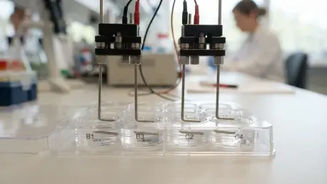

Electronic controller modules based on Arduino firmware now allow for the simultaneous testing of multiple electrical pulse protocols within a single standard 12-well plate. This breakthrough by researchers at the Institute of Life and Health Sciences in Portugal addresses one of the most persistent bottlenecks in modern regenerative medicine: the inability to efficiently replicate the complex bioelectric environment of the human body in a controlled laboratory setting. While cells naturally respond to electrical gradients during development and wound healing, traditional tools for studying these phenomena have often been bulky, expensive, and limited to testing one variable at a time. This new multi-channel in vitro electrical stimulator represents a paradigm shift, providing a scalable platform that integrates seamlessly with existing laboratory workflows. By enabling high-throughput experimentation, the system allows scientists to observe how various cell types react to diverse electrical intensities and frequencies in parallel, significantly reducing the time required to identify optimal therapeutic parameters. The democratization of such technology is vital as the scientific community seeks to move beyond chemical-only treatments, looking instead toward the “electrome” to solve complex neurological and musculoskeletal challenges.
Engineering a Simplified Stimulation Interface
The structural design of this new stimulator prioritizes functional simplicity without sacrificing the precision required for high-stakes biomedical research. The primary interface consists of a custom-engineered lid fabricated from 5 mm thick acrylic, which serves as a stable mounting platform for the electrical components. Attached to this lid are L-shaped electrodes meticulously crafted from platinized titanium foil. This specific configuration was selected because it allows for easy assembly and cleaning, which is essential for maintaining sterile conditions during long-term cell culture experiments. Unlike previous generations of stimulators that relied on fragile or difficult-to-manufacture components, this design utilizes widely available materials that can be easily replaced or modified. The use of a 12-well plate format ensures that the device is compatible with standard imaging and analytical tools already present in most biology labs, making it an accessible upgrade for researchers exploring the frontier of electrophysiology.
The selection of platinized titanium for the electrodes is a deliberate choice intended to solve long-standing issues regarding electrochemical stability and biological safety. Titanium provides a robust and cost-effective substrate that resists mechanical deformation, while the platinum coating facilitates a stable flow of current through the culture media without the risk of heavy metal leaching or electrode degradation. A significant technical advancement in this setup is the direct placement of electrodes into the culture wells, which completely bypasses the need for traditional salt-bridges. Salt-bridges, though common in older setups, are notoriously problematic because they are prone to clogging, evaporation, and the formation of air bubbles that can interrupt electrical continuity and ruin weeks of experimental progress. By employing alternating current pulses, the Portuguese research team has successfully managed the potential for reactive oxygen species or pH shifts, ensuring that the biological outcomes observed are a direct result of the electric field rather than toxic byproducts of the stimulation process.
Precision Mapping and Modeling of Electric Fields
To ensure that the electrical signals delivered to the cells are both accurate and reproducible, the researchers utilized advanced finite element modeling to visualize the electric field distribution within each individual well. This computational approach allowed the team to define a “stimulation zone”—a specific rectangular area at the center of each well where the electric field remains within a strict 10% tolerance of the intended strength. Identifying this zone is critical for scientific validity, as it ensures that cells located in different parts of the well are not subjected to vastly different electrical forces. Without such precision, data collected from high-throughput plates would be marred by inconsistencies, making it nearly impossible to draw meaningful conclusions about the efficacy of a particular pulse protocol. This rigorous mapping provides a standardized framework that allows other laboratories to replicate these findings with a high degree of confidence, bridging the gap between theoretical physics and practical biology.
The simulations also yielded critical insights into how the physical environment of the culture well influences electrical intensity, specifically identifying an inverse relationship between media volume and electric field strength. This means that even a slight inaccuracy in the amount of liquid added to a well could significantly alter the dose of electricity a cell receives, highlighting the necessity for meticulous pipetting in electro-stimulation experiments. Interestingly, the model demonstrated that the vertical position of the electrodes—how high they sit from the bottom of the well—has a negligible impact on the field strength, provided they are sufficiently submerged. This discovery provides researchers with a degree of physical flexibility when setting up their plates, allowing for variations in cell culture height without compromising the integrity of the electric field. Such technical clarity is essential for developing robust protocols that can survive the transition from a specialized physics laboratory to a broader biological research context.
Advanced Control and Modular Programming
At the heart of the system lies a custom-built modular controller powered by sophisticated Arduino firmware, which acts as the central command unit for the entire 12-well array. This “electronic brain” is constructed using stacked, two-layer printed circuit boards that allow for independent control over each well, enabling the simultaneous execution of twelve different stimulation protocols on a single plate. One of the most significant engineering choices in this controller is its operation in constant current mode. Traditional constant voltage systems often suffer from fluctuations in the electric field because the electrical resistance of the media and the electrodes changes over time as cells grow and nutrients are consumed. By maintaining a constant current, the device ensures that the electrical pulses remain perfectly rectangular and the amplitudes remain precise throughout the duration of the experiment, regardless of the shifting electrochemical environment within the well.
The flexibility of the firmware allows researchers to program a wide array of stimulation parameters, including the specific duration of each pulse, the amplitude of the signal, and the frequency of the stimulation cycles. This programmable versatility is what makes the tool so powerful for comparative studies, as it allows scientists to rapidly screen a broad spectrum of intensities—up to 160 V/m—to see which specific settings trigger the desired cellular response. For instance, a researcher could test high-frequency signals modeled after spinal cord stimulation alongside low-frequency signals intended to mimic natural nerve growth, all within the same experimental run. This capacity for parallel processing not only accelerates the pace of discovery but also ensures that all variables are tested under identical environmental conditions, such as temperature and CO2 levels, thereby eliminating many of the external factors that can lead to experimental bias.
Biological Impacts on Neuronal Development
The efficacy of this multi-channel tool was rigorously validated through its application to human neuroblastoma cells and neuronal progenitor cells, revealing profound effects on physical growth and maturation. In experiments using the SH-SY5Y cell line, the team applied various protocols to observe how electricity influences neurite outgrowth—the critical process where neurons sprout the branches necessary for communication. The results indicated that certain high-frequency and high-amplitude settings significantly boosted the length and complexity of these branches compared to non-stimulated controls. Importantly, subsequent metabolic assays confirmed that these bursts of electricity did not harm the cells, proving that the accelerated growth was a healthy physiological reaction. This finding is particularly relevant for treating nerve injuries, as it suggests that targeted electrical pulses could be used to guide and speed up the repair of damaged neural pathways in a clinical setting.
Further exploration into human neuronal progenitor cells showed that electrical stimulation could also act as a powerful driver for differentiation, essentially telling immature cells to “grow up” into functional neurons. A fascinating aspect of this research was the discovery of how electrical signals interact with existing chemical growth factors. The data suggested that when chemical cues were absent, the impact of the electric field was much more pronounced, whereas the presence of chemicals seemed to saturate the cell’s developmental pathways. This suggests that “electroceuticals”—medical treatments using electrical signals—might work through similar biological mechanisms as traditional drugs, offering a new way to stimulate tissue regeneration without the side effects often associated with systemic chemical treatments. The ability to precisely tune these signals using the new stimulator lid provides a pathway for developing personalized electrical therapies tailored to a patient’s specific cellular needs.
Exploring Complex Cultures and Stem Cell Secretions
The application of the stimulator to primary spinal cord cells from rat models provided a more complex look at how electricity affects diverse cell populations within a single environment. In these mixed cultures, the electrical pulses not only encouraged neuronal growth but also influenced the behavior of surrounding glial cells, which are the support structures for the nervous system. However, the study also highlighted a critical “therapeutic window,” as certain high-intensity settings actually led to a decrease in the overall percentage of neurons. The researchers hypothesized that this was a result of excitotoxicity, where excessive electrical activity causes a cell to become overstimulated and eventually die. This discovery is a vital cautionary tale for the field of bioelectronics, emphasizing that more stimulation is not always better and that the multi-channel nature of this tool is essential for finding the delicate balance between promoting healing and causing harm.
Perhaps the most groundbreaking discovery involved the use of human adipose-derived stem cells, where the electricity was used not to change the cells themselves, but to alter their “secretome.” The secretome is the cocktail of proteins, growth factors, and signaling molecules that stem cells release into their surroundings to communicate with other cells. By applying specific electrical protocols, the researchers were able to “prime” these stem cells, causing them to release a more potent mix of factors that helped other progenitor cells differentiate into mature neurons. This indicates that electrical stimulation can be used to turn stem cells into biological factories for repair, producing customized therapeutic fluids that can be collected and used to treat neurological conditions. This indirect method of therapy—using electricity to enhance the natural healing power of stem cells—represents a significant new frontier in regenerative medicine that could lead to more effective treatments for chronic degenerative diseases.
Technical Validation and Future Collaborative Implementation
To ensure the long-term reliability of the stimulation platform, the research team conducted exhaustive safety tests, monitoring the physical stability of the culture environment during extended periods of use. Using high-precision PT 1000 thermal sensors, they verified that the electrical pulses did not cause any significant increase in the temperature of the culture media, which is a common failure point for cheaper, less refined stimulation devices. Additionally, continuous pH monitoring confirmed that the alternating current protocols effectively prevented the accumulation of toxic electrolytic byproducts, maintaining a stable and healthy environment for the cells. These findings established that the device is safe for the multi-day experiments required to observe complex biological processes like stem cell differentiation or tissue scaffolding, providing a level of technical assurance that is often missing from homemade laboratory equipment.
While the current results are promising, the next logical steps for this technology involved moving toward more advanced functional testing to confirm the maturity of the stimulated cells. Future studies planned for the 2027 to 2029 period were expected to incorporate patch-clamp electrophysiology to see if these electricity-grown neurons can fire action potentials like their naturally occurring counterparts. The open-access design of the system, coupled with its reliance on modular components, encouraged other institutions to adopt and adapt the lid for their own specific research needs, such as muscle tissue engineering or bone regeneration. Researchers were encouraged to experiment with different electrode geometries and conductive hydrogels to further refine the interface between hardware and biology. By providing a blueprint for accessible, high-throughput stimulation, the team from Portugal moved the entire field closer to a future where electrical medicine is as common and well-understood as the pharmaceutical industry.