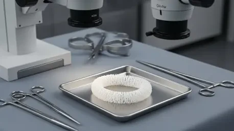

Trauma surgeons often face a race against time when a patient presents with a shattered limb or a ruptured artery where every second spent searching for a viable donor vein increases the risk of amputation or death. This high-pressure environment has long necessitated a surgical solution that transcends the physical limitations of existing grafting methods. The recent emergence of the Human Acellular Vessel (HAV) offers a glimpse into a new standard of care where bioengineered, shelf-ready tissues are available for immediate implantation. Unlike previous biological attempts, these vessels are meticulously designed to be immunologically neutral while providing the structural integrity required for high-pressure arterial flow. This development marks a transition away from autologous harvesting, where surgeons must create a second wound to repair the first. By integrating these lab-grown scaffolds into clinical practice, medical professionals are beginning to address the critical shortage of viable vascular material. The ability to store these vessels in a standard refrigerator ensures that life-saving interventions are not delayed by biological availability or complex preparation protocols.
Overcoming Limitations of Traditional Grafting Techniques
For several decades, the surgical community viewed the autologous graft as the gold standard for restoring blood flow in patients with damaged or diseased arteries. This process typically involves harvesting the saphenous vein from the patient’s leg to bypass or repair a blockage elsewhere in the body. While this method uses the patient’s own tissue, it is inherently flawed because it requires a second, often invasive, surgical site that significantly increases recovery time and the potential for wound complications. Furthermore, many patients who require vascular intervention suffer from co-morbidities such as advanced diabetes, obesity, or peripheral artery disease, which can render their own veins unsuitable for use. This vein depletion often leaves surgeons with no high-quality biological options, forcing them to turn to less effective alternatives that may not hold up over time. The reliance on the patient’s own limited biological resources has historically capped the success rate of complex revascularization procedures in the most vulnerable populations.
When a patient’s own veins are unavailable, the secondary alternative has traditionally been the use of synthetic plastic grafts made from materials like polytetrafluoroethylene or Dacron. Although these manufactured tubes are easy to store and come in various sizes, they function as foreign objects within the human body and lack the ability to integrate with native tissue. Because these synthetic materials do not possess a cellular component, they cannot grow, heal, or respond to physiological signals in the way a natural artery does. This biological disconnect makes them highly susceptible to chronic infections that are notoriously difficult to treat, often requiring the complete removal of the graft. Over time, the lack of endothelization on the inner surface of synthetic grafts also increases the risk of thrombosis, leading to sudden failure and the need for repeat interventions. The persistent danger of infection and the mechanical mismatch between rigid plastic and flexible human tissue have long highlighted the urgent need for a more biologically compatible, living alternative for vascular surgery.
Engineering Resilience Through Decellularized Scaffolds
The development of bioengineered vessels begins in a highly controlled laboratory setting where donor muscle cells are seeded onto a specialized biodegradable scaffold. These cells are placed into a bioreactor that subjects the nascent tissue to cyclical mechanical pressure, effectively exercising the vessel to simulate the pulse and flow of a human circulatory system. This mechanical stimulation is essential because it encourages the cells to produce a dense, organized matrix of collagen and other structural proteins that provide the graft with its necessary strength and elasticity. Over several weeks, the scaffold dissolves as the biological matrix matures, resulting in a tube that closely mimics the physical properties of a native human artery. This bio-mimetic approach ensures that the resulting vessel can withstand the intense physiological pressures of arterial blood flow without dilating or rupturing. By focusing on the structural foundations of human biology, engineers have created a material that is both robust enough for surgical handling and flexible enough to integrate into the moving environment of the human body.
Once the structural matrix is complete, the vessel undergoes a rigorous decellularization process that removes all donor cellular material while leaving the protein-based skeleton intact. This step is critical because it eliminates the antigens that would otherwise trigger an immune rejection from the recipient, allowing the vessel to be used universally across different patients without the need for matching. The remaining acellular scaffold is essentially a biological chassis that serves as a guide for the recipient’s own cells once it is implanted into the body. Following surgery, the patient’s native cells begin to migrate into the protein matrix, gradually transforming the inanimate graft into living tissue that the body recognizes as its own. This host remodeling process allows the vessel to develop a functional inner lining, which is vital for preventing blood clots and resisting infection. Unlike synthetic tubes, this bioengineered tissue becomes a dynamic part of the patient’s anatomy, capable of self-repair and metabolic interaction. This transition from an engineered product to a living biological entity represents a fundamental advancement in regenerative medicine.
Scaling Bioengineered Solutions for Complex Procedures
While initial clinical applications have focused on larger vessels for treating traumatic limb injuries, the research trajectory starting in 2026 is moving toward scaling this technology for even more demanding procedures. Engineers are currently refining the manufacturing process to produce vessels with diameters as small as two to three millimeters, which are necessary for coronary artery bypass grafting. This move toward smaller scales is technically challenging, as the vessels must maintain their structural integrity and patency in the low-flow, high-resistance environment of the heart’s surface. Successfully implementing small-diameter bioengineered grafts could revolutionize the treatment of cardiovascular disease by providing an endless supply of high-quality bypass material. This shift reduces the surgical complexity of heart surgery by eliminating the need to harvest veins from the chest or legs, thereby reducing operating times and improving patient outcomes. As these smaller vessels move through clinical evaluations from 2026 to 2028, the potential for a new era in cardiac care becomes increasingly tangible, moving the field away from purely mechanical or pharmacological management.
The evolution of these advanced materials reflects a broader shift toward a third wave of medical therapy that utilizes engineered human tissues as a universal tool for repair. Beyond vascular surgery, the underlying platform of decellularized protein scaffolds offers potential applications in repairing other tubular structures in the body, such as the urinary tract or esophageal tissues. By bridging the gap between biological science and industrial engineering, medical researchers are no longer limited by the scarcity of donor organs or the biological incompatibility of plastics. The focus has moved from creating isolated medical devices to establishing a versatile regenerative framework that can be adapted for a wide variety of diseases and traumatic conditions. This integrated approach allows for the mass production of biological tissues that can be shipped globally, ensuring that advanced surgical options are available to patients regardless of their location. As the technology matures, the distinction between a laboratory-grown product and a natural body part will continue to blur, paving the way for a future where damaged anatomy is replaced with tissue that is functionally indistinguishable from the original.
Advancing the Standards of Bioengineered Vascular Intervention
The implementation of bioengineered vessels provided a definitive answer to the long-standing challenges of vascular reconstruction and emergency revascularization. Medical facilities that integrated these shelf-ready tissues observed a marked decrease in the complications associated with traditional autologous harvesting and synthetic grafting failures. By prioritizing biological compatibility and immediate availability, surgical teams minimized the time spent in the operating room while maximizing the long-term success rates of arterial repairs. The transition toward decellularized scaffolds encouraged a more proactive approach to patient care, where the limitations of a patient’s own donor material no longer dictated the quality of the intervention. Researchers focused on refining the host remodeling process, ensuring that the integration of these vessels into the circulatory system remained consistent across diverse patient populations. This strategic shift in surgical practice emphasized the importance of regenerative materials as a primary tool for restoring human health. The progress made in bioengineering eventually established a new baseline for surgical excellence, where the focus remained on providing durable, infection-resistant solutions for complex vascular conditions.