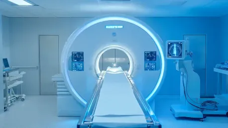
In the midst of technological advancements, medical imaging tests like X-rays, MRIs, mammograms, and CT scans have become indispensable tools in modern diagnostics; however, questions about the safety and health risks associated with these tests frequently emerge. Radiation exposure is a common concern, with varying levels emitted depending on the type of imaging test. To alleviate these concerns, it is essential to understand the nuances of radiation risks, guidelines, and the importance of patient transparency regarding their medical imaging history.
Radiation Exposure Levels
Different imaging tests emit varying levels of radiation, with some posing minimal risk and others requiring more careful consideration. X-rays, bone density scans, and mammograms typically involve lower levels of radiation, comparable to a few days of background radiation from natural sources like the ground, air, or cosmic rays. For instance, a simple X-ray carries a negligible risk, making it a routine diagnostic tool. In contrast, higher radiation exposures from CT or PET scans still pose a low risk of cancer, but they are roughly equivalent to years of natural radiation exposure. Understanding these distinctions helps patients and healthcare providers make informed decisions about the necessity and frequency of such tests.
Frequency of Scans
There is no strict upper limit on the number of imaging tests a patient can undergo in a year, as the decision largely depends on the patient’s medical conditions and history. It is crucial for patients to inform their doctors of any recent scans, particularly those performed in private facilities or overseas, to prevent unnecessary repetition. This transparency not only minimizes repeated radiation exposure but also ensures that the healthcare provider has a comprehensive understanding of the patient’s current medical situation, facilitating better diagnosis and treatment planning.
Reasons for Multiple Imaging Tests
Different imaging modalities often complement each other to provide a comprehensive view of a patient’s condition. For example, while X-rays are effective in detecting bone fractures, they might need to be supplemented with CT or MRI scans to check for organ damage or internal bleeding. Utilizing multiple imaging methods ensures precise diagnosis, effective treatment planning, and thorough monitoring of disease progression. Each modality offers unique insights that, when combined, contribute to a holistic understanding of the patient’s health.
Dental X-Rays
Dental X-rays are generally safe, but there are specific considerations to keep in mind. Pregnant women should notify their dentist before undergoing any X-rays due to potential risks to the fetus. However, modern dental X-ray equipment is designed to minimize radiation exposure to only the targeted area, often making additional protective measures like thyroid collars unnecessary. This balance between safety and diagnostic efficacy enables the continued use of dental X-rays in managing oral health without significant risk.
Specific Imaging Tests
X-Rays
X-rays are suitable for detecting fractures and infections with low radiation doses, making them a common and accessible diagnostic tool. However, pregnant women should avoid X-rays due to potential risks to the fetus. Despite this, X-rays remain a vital component of medical diagnostics due to their simplicity and effectiveness in identifying various conditions.
CT Scans
CT scans provide detailed visualization of internal organs and are often used when more intricate imaging is required. These scans may involve a contrasting agent, which could be unsuitable for individuals with asthma, allergies, or renal impairment. Despite the higher radiation exposure, the detailed insights gained from CT scans are invaluable for complex medical evaluations.
MRI
MRIs use magnetic fields rather than radiation, making them a safer option for young adults and pregnant women. However, certain contraindications like metallic implants and claustrophobia can limit their use. MRIs are particularly useful for soft tissue imaging, offering detailed views that are crucial in various medical scenarios.
Ultrasound
Ultrasound imaging is safe for use during pregnancy and involves no radiation, making it a preferred choice for fetal monitoring and other applications. While effective for many diagnostic purposes, ultrasounds are less useful for imaging bone and gas-filled areas, limiting their scope in certain situations.
Mammograms
Mammograms are specifically used for breast screening and are not recommended for younger women until they reach the appropriate screening age due to radiation exposure. Despite this, mammograms play a crucial role in early breast cancer detection, significantly contributing to effective treatment.
Bone Density Scans
Bone density scans assess bone strength, typically used for older adults or individuals on certain medications. These scans, however, are not performed on pregnant women due to potential risks. By evaluating bone density, these scans help in diagnosing conditions like osteoporosis and planning appropriate treatments.
PET Scans
PET scans are specialized for detecting cancer spread and require prolonged scanning periods due to the absorption time of the radioactive dye. Generally avoided in children and pregnant women, PET scans provide critical information for cancer diagnosis and treatment planning, despite the specific precautions needed.
Trends and Overarching Views
The primary consensus is that while all imaging techniques carry some radiation risk, these risks are generally low and justified by the substantial benefits in accurate diagnosis and effective treatment planning. Emphasizing patient transparency about scan history minimizes unnecessary repeated exposure. Overall, the advantages of these imaging tests in ensuring accurate medical diagnoses significantly outweigh the minimal associated risks.
Summary
In the era of rapid technological progress, medical imaging tests like X-rays, MRIs, mammograms, and CT scans have become vital tools for modern diagnostics. Despite their value, concerns about their safety and potential health risks often arise, particularly regarding radiation exposure. The level of radiation risk changes with each type of imaging test, and understanding these differences is crucial for alleviating anxiety. It’s vital to grasp the intricacies of radiation risks and follow established guidelines to ensure patient safety. Additionally, transparency about a patient’s medical imaging history is important for both patients and healthcare providers. This knowledge helps in making informed decisions about the necessity and frequency of these tests. By being aware of the advantages and risks, healthcare providers can use these indispensable diagnostic tools more effectively while prioritizing patient health. Ensuring that patients are well-informed about the risks and benefits of their imaging tests is a critical component of modern medical practice.


