
In emergency rooms, prenatal clinics, and primary care offices alike, clinicians reach for ultrasound because it delivers immediate answers without exposing patients to ionizing radiation, yet it still runs through the same radiology workflows that govern X-rays and CT. That dual identity raises a practical question with financial stakes: is ultrasound truly “radiology,” and what does that mean for billing, coverage, and out-of-pocket costs? The answer threads through how the technology operates, why it has become central to modern care, and how health plans adjudicate medical necessity. Understanding that landscape helps patients plan, avoid surprise bills, and get the most from a test designed to be safe, fast, and remarkably versatile across settings.
What Ultrasound Is and Where It Fits in Radiology
Ultrasound is diagnostic imaging built on sound, not radiation: a transducer sends high-frequency waves into the body, gel improves transmission, and returning echoes are converted into real-time images of organs, vessels, and motion. This physics separates it from radiographic modalities, yet operationally it sits squarely within diagnostic radiology. Sonographers acquire images using standardized protocols; radiologists or qualified clinicians interpret those images and issue reports that guide treatment. That workflow aligns with how hospitals and imaging centers staff, schedule, and document studies, so the service is organized and billed under radiology even when used at the bedside or in clinics. The classification reflects care delivery and quality assurance rather than the energy source.
Ultrasound’s placement in radiology also mirrors coding and compliance rules that underpin payment. Most studies are identified by CPT codes that separate the technical component (equipment, room time, technologist) from the professional component (interpretation), and many facilities submit them as distinct claims. This structure supports peer review, report archiving, and integration with electronic health records. It also anchors accreditation standards for equipment performance and operator competency, which protect diagnostic quality despite wide variation in practice settings. Therefore, while lay readers may equate “radiology” with radiation, the term in healthcare broadly denotes the domain of diagnostic imaging services, within which ultrasound is a core modality.
How Ultrasound Works and What It’s Used For
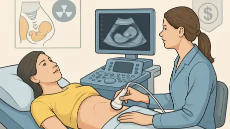
At its core, ultrasound measures how sound waves reflect differently from fluid, soft tissue, fat, and more solid structures. The system converts these echoes into gray-scale images; when Doppler is applied, it visualizes and quantifies blood flow using color or spectral traces. Because this all happens in real time, clinicians can watch valves open, track fetal movement, or guide a needle tip to a target as it advances. That immediacy supports focused questions: is there gallbladder inflammation, a deep vein thrombosis, or a pericardial effusion? With modern machines, image quality and Doppler sensitivity have improved, expanding what can be answered at the point of care.
Clinical applications span the abdomen, pelvis, thyroid, breast, vascular system, and heart, along with musculoskeletal and procedural guidance. Echocardiography assesses chamber size and function, Doppler evaluates stenoses and venous clots, and obstetric ultrasound monitors fetal growth, placental position, and well-being. The modality excels at soft-tissue characterization that would be invisible on plain films, yet it remains operator dependent: outcomes hinge on probe selection, patient positioning, and the sonographer’s ability to optimize the acoustic window. Body habitus and overlying gas can obscure views, and deep or air-filled structures may require CT or MRI. Used for the right indication, however, ultrasound frequently delivers decisive, actionable findings.
Safety, Strengths, and Limits in Modern Care
A consistent advantage is safety: because ultrasound uses sound waves, there is no ionizing radiation exposure to accumulate over a lifetime. That makes it first-line for children and pregnant patients, and suitable for repeat assessments when conditions evolve. Beyond the absence of radiation, exams are noninvasive, typically painless, and quick to schedule. Portability pushes the value further. Compact systems travel to the bedside, rural clinics, and even ambulances, where time-sensitive decisions benefit from immediate imaging. Professional societies and global health agencies endorse responsible use in these settings, citing gains in diagnostic accuracy and triage efficiency without sacrificing safety.
Nevertheless, users must respect limits. Thermal and mechanical indices displayed on modern scanners guide safe output settings, and trained operators adjust exposure to the lowest level necessary to achieve the diagnostic goal. Image quality also varies with equipment, patient anatomy, and the clinical question; a state-of-the-art system cannot rescue an unfocused exam or a question poorly matched to ultrasound’s strengths. In practice, ultrasound complements rather than replaces CT and MRI, often narrowing the problem quickly and determining whether further imaging is required. This tiered approach preserves speed and safety while ensuring comprehensive evaluation when complex or deep-seated disease is suspected.
Billing, Coverage, and Costs in the United States
For billing, ultrasound is managed as radiology. The typical claim includes a technical fee for the exam itself and a professional fee for interpretation, sometimes from separate entities that must both be in network to avoid balance billing. Coverage hinges on medical necessity: studies ordered to evaluate symptoms, abnormal labs, or known conditions are generally covered under diagnostic imaging benefits. Elective keepsake scans, including non-medical gender-reveal imaging, are usually excluded. Obstetric baselines are commonly covered, while additional targeted studies may require prior authorization. Plans also set rules around site of care, steering patients to contracted facilities and credentialed readers.
Out-of-pocket amounts vary by plan design and timing. With insurance, many patients see copays or coinsurance in the ballpark of $20 to $200, with higher costs if a deductible has not been met. Without insurance, self-pay prices often land between $150 and $600, though specific exams cluster within narrower bands—abdominal studies around $200 to $400 and pelvic studies about $250 to $500 are common reference points. Hospitals tend to post higher rates than independent imaging centers because of overhead and facility fees, and regional pricing differences can be significant. Transparency tools from insurers and public databases help forecast costs, but confirmation with the imaging provider remains the most reliable step.
Practical Steps and Closing Insights
Financially, patients can tilt the odds in their favor by comparing sites before scheduling, asking about bundled cash rates, and confirming that both the facility and the interpreting radiologist are in network. Independent imaging centers frequently offer lower prices for the same CPT-coded exam, and many provide self-pay discounts when payment is made at the time of service. Pre-service estimates reduce surprises, as does clarifying whether Doppler or extended protocols might be added during the visit. When a second opinion is appropriate, remote consultation can deliver subspecialty expertise without duplicating the exam. Preparation matters, too: fasting for 6–12 hours improves abdominal imaging, a full bladder enhances pelvic views, and most thyroid or breast scans require no special steps.
Taken together, the evidence showed that ultrasound belonged within radiology’s operational universe while remaining distinct in physics, and that its lack of ionizing radiation, real-time capabilities, and portability made it indispensable across care settings. Costs were shaped more by site of care and insurance design than by the test itself, suggesting that proactive planning, network checks, and price comparisons yielded meaningful savings. Simple preparation steps increased diagnostic yield, and recognition of limitations helped clinicians decide when to escalate to CT or MRI. As devices continued to shrink and image quality improved, ultrasound’s role in timely, patient-centered decision-making had only grown more central.