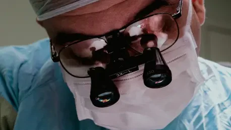
Glioblastoma, recognized as the most aggressive and deadly form of brain cancer, presents an immense challenge for neurosurgeons tasked with removing tumors that infiltrate healthy brain tissue with devastating precision. The difficulty in achieving complete resection without impairing critical brain functions often results in residual cancer cells, paving the way for recurrence and poor patient outcomes. A remarkable breakthrough comes in the form of an optical imaging probe developed through a collaboration between Erasmus University Medical Center in the Netherlands and the University of Missouri in the USA. This cutting-edge tool promises to revolutionize surgical approaches by enabling real-time detection of microscopic cancer cells at tumor margins, offering a potential lifeline for improved safety and efficacy during these high-stakes procedures. By addressing a core issue in glioblastoma treatment, this innovation sparks hope for better survival rates and quality of life for patients battling this formidable disease.
Advancements in Surgical Imaging Technology
Targeting Metabolic Vulnerabilities
The innovative optical probe, dubbed FA-ICG, represents a significant leap forward by merging a long-chain saturated fatty acid with indocyanine green (ICG), a near-infrared dye already approved for clinical applications. This strategic combination exploits the heightened fatty acid (FA) metabolism inherent to glioblastoma cells, a crucial driver of their rapid proliferation and invasiveness. By accumulating preferentially in tumor tissues, the probe emits a potent fluorescence signal that sharply delineates cancerous areas from surrounding healthy brain matter. The use of ICG enhances this capability, offering deep tissue penetration and a superior signal-to-noise ratio, which are vital for clear visualization during surgery. This targeted approach not only aids in identifying tumor boundaries but also minimizes the risk of overlooking minute cancer clusters, potentially transforming the precision of neurosurgical interventions.
Beyond its biochemical ingenuity, the FA-ICG probe stands out for its adaptability to the operating room environment, aligning seamlessly with existing surgical imaging technologies. Its design ensures that surgeons can rely on familiar near-infrared systems to detect the fluorescence signals without needing extensive retraining or equipment overhauls. The metabolic focus on FA uptake also opens doors to understanding glioblastoma behavior at a cellular level, providing insights that could inform future therapeutic strategies. As a non-invasive imaging tool, it reduces the need for additional invasive procedures to confirm tumor margins, thereby decreasing patient risk during already complex surgeries. The specificity of this probe marks a pivotal shift in how metabolic traits of cancer can be harnessed for practical medical advancements, setting a new standard for intraoperative visualization and decision-making.
Harnessing Visual Clarity for Surgical Success
The clarity provided by the FA-ICG probe during surgical procedures offers a transformative advantage, allowing neurosurgeons to navigate the intricate landscape of the brain with unprecedented accuracy. Unlike traditional imaging methods that often struggle to differentiate subtle tumor margins, this probe’s fluorescence signals illuminate even the smallest clusters of glioblastoma cells in real time. This heightened visibility is critical in a disease where every remaining cancer cell can contribute to recurrence, significantly impacting long-term patient prognosis. Preclinical data highlights how this tool enhances a surgeon’s ability to make informed decisions on the spot, potentially reducing the duration of surgeries by streamlining the identification process. Such efficiency could lower the physical and emotional toll on patients undergoing these grueling operations.
Additionally, the probe’s integration of near-infrared technology ensures that its benefits are not hampered by the challenges of deep-seated tumors, a common obstacle in brain cancer surgeries. The ability to penetrate tissue depths while maintaining signal strength means that even tumors located in hard-to-reach areas can be accurately mapped out for resection. This addresses a long-standing limitation in glioblastoma treatment, where incomplete removal due to poor visibility often necessitates follow-up interventions. The FA-ICG probe also holds promise for reducing collateral damage to healthy brain tissue, preserving neurological functions that are often compromised during aggressive tumor removal. By providing a clearer picture of the surgical field, this technology empowers medical teams to balance the dual goals of thorough cancer eradication and patient safety, potentially redefining success metrics in neurosurgery.
Preclinical Success and Practical Applications
Validation Through Rigorous Testing
Extensive preclinical evaluations have laid a robust foundation for the FA-ICG probe, demonstrating its efficacy in controlled settings that mirror real-world challenges. Initial cellular studies confirmed that the probe effectively replicates the natural uptake of fatty acids in living glioblastoma cells, ensuring its biological relevance. Subsequent trials in mice implanted with glioblastoma tumors revealed fluorescence signals approximately 2.2 times stronger than those produced by standalone ICG, underscoring the probe’s superior targeting capability. Moreover, retention rates in tumor-bearing brain regions were markedly higher, affirming its specificity for cancerous tissues over healthy ones. These findings suggest that the probe could significantly enhance the ability to detect residual cancer cells that might otherwise evade traditional imaging, offering a critical edge in preventing tumor regrowth.
Further testing in patient-derived glioblastoma models has illuminated additional benefits, particularly in tracking tumor progression over extended periods. This capability proves invaluable for research purposes, as glioblastoma often exhibits unpredictable growth patterns that complicate preclinical studies. The probe’s consistent performance across diverse experimental setups highlights its reliability as a diagnostic and monitoring tool, providing a clearer understanding of disease dynamics. Such insights are essential for developing complementary therapies that can work in tandem with surgical interventions to improve patient outcomes. The rigorous validation process not only builds confidence in the probe’s potential but also sets a high benchmark for future innovations in cancer imaging, pushing the boundaries of what is possible in precision medicine.
Real-World Surgical Guidance
The practical utility of the FA-ICG probe shines through in its application to fluorescence-guided surgery, as demonstrated in preclinical trials with tumor-bearing mice. Using standard near-infrared cameras already approved for surgical use, the probe delivered significantly stronger fluorescence signals compared to ICG alone, enabling precise visualization of tumor margins during operations. This enhanced clarity allowed for more accurate resections, addressing the persistent challenge of incomplete tumor removal in glioblastoma cases. The ability to integrate with existing surgical infrastructure without requiring extensive modifications further bolsters its appeal, ensuring that hospitals can adopt this technology with minimal disruption to current practices. Such seamless implementation is crucial for widespread acceptance in high-pressure clinical environments.
Beyond rodent models, the probe’s versatility was showcased in a veterinary setting during the surgical resection of a canine mastocytoma, a type of skin cancer in dogs. Ten hours post-injection, the FA-ICG probe facilitated successful image-guided surgery using an open-air near-infrared camera, proving its adaptability across different species and cancer types. This cross-species efficacy hints at broader applications in surgical oncology, potentially benefiting a wider range of patients and conditions. The real-world implications of these findings are profound, suggesting that the probe could serve as a universal tool for enhancing surgical precision in various contexts. As a result, it lays the groundwork for innovative approaches to cancer treatment, bridging the gap between experimental success and tangible clinical impact.
Future Prospects and Clinical Potential
Path to Human Trials
As the FA-ICG probe moves closer to clinical application, the research team is diligently preparing for a Phase I clinical trial to assess its safety, tolerability, and effectiveness in human patients facing glioblastoma. This crucial step will determine whether the promising preclinical results translate into real benefits for individuals undergoing brain cancer surgery. The probe’s compatibility with existing surgical cameras and microscopes, along with its suitable half-life and visibility under standard operating room lighting, positions it as a practical addition to current medical protocols. Successful outcomes from these trials could herald a new era in neurosurgery, where enhanced tumor visualization becomes a routine part of treatment plans, ultimately improving the efficacy of subsequent therapies like chemotherapy and radiation by ensuring more comprehensive tumor removal.
The journey to human trials also underscores the importance of addressing potential hurdles, such as regulatory approvals and scalability for widespread clinical use. Ensuring that the probe meets stringent safety standards is paramount, as is demonstrating its cost-effectiveness for healthcare systems under financial strain. Collaboration between researchers, clinicians, and industry stakeholders will be essential to navigate these challenges and bring the technology to market. If the trials confirm the probe’s benefits, it could pave the way for accelerated adoption in hospitals worldwide, offering a lifeline to countless patients. The anticipation surrounding this phase reflects a broader optimism within the medical community about the role of advanced imaging in transforming cancer care, highlighting the probe’s potential to set a new benchmark for surgical innovation.
Shaping the Future of Cancer Surgery
Reflecting on the strides made with the FA-ICG probe, it’s evident that past efforts in preclinical and veterinary settings yielded encouraging outcomes that reshaped expectations for glioblastoma treatment. The probe’s ability to highlight microscopic cancer cells in real time during surgery marked a significant departure from traditional methods, providing neurosurgeons with a tool to minimize damage to healthy tissue while maximizing tumor removal. Each test, from cellular studies to complex surgical applications, reinforced the probe’s specificity and imaging strength, offering a glimpse into a future where precision defined every cut. The medical field watched closely as these developments unfolded, recognizing the potential to delay cancer recurrence through more thorough interventions.
Looking ahead, the focus shifted to actionable steps following the completion of initial trials. Expanding research to include diverse patient populations emerged as a priority to ensure the probe’s efficacy across different demographics. Additionally, integrating this technology with emerging digital surgery platforms could further enhance its precision, creating a synergy that benefits both surgeons and patients. The momentum built from past successes suggested that continued investment in fluorescence-guided surgery would likely yield even greater advancements, potentially extending the probe’s utility to other aggressive cancers. These next steps promised to build on the foundation laid by earlier achievements, driving forward a vision of safer, more effective cancer surgeries.


