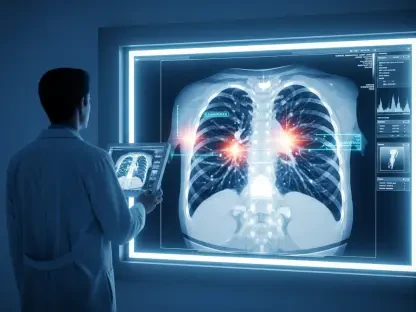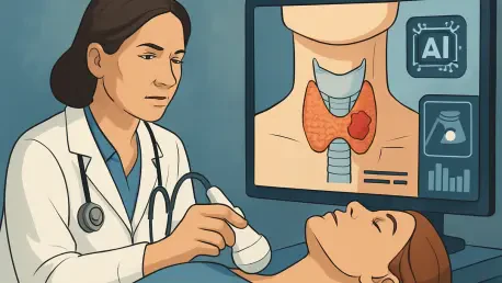

Imagine a diagnostic tool so advanced that it can uncover hidden threats within the thyroid gland, even when masked by a complex inflammatory condition. A groundbreaking study has brought this vision to life by developing a radiomics model based on nonenhanced computed tomography (NECT) scans to detect papillary thyroid carcinoma (PTC) in patients with Hashimoto’s thyroiditis (HT). This common autoimmune disorder often obscures the presence of malignant changes due to chronic inflammation, posing a significant challenge for early diagnosis. The research, recently published in a leading medical journal, introduces a novel approach that leverages machine learning and detailed imaging analysis to address this critical gap. By offering a non-invasive way to identify PTC amidst the diffuse changes of HT, this innovation holds the promise of transforming thyroid cancer detection, ensuring timely intervention for a population at heightened risk. This development not only tackles a persistent clinical hurdle but also paves the way for broader applications in medical imaging, potentially benefiting countless patients worldwide.
Advances in Thyroid Cancer Detection
Challenges in Diagnosing PTC in HT
Inflammatory Background Complications
Detecting papillary thyroid carcinoma in the context of Hashimoto’s thyroiditis presents a formidable obstacle for clinicians due to the overlapping characteristics of these conditions on standard imaging. Hashimoto’s thyroiditis, marked by chronic inflammation and lymphocytic infiltration, creates a diffuse, heterogeneous appearance in the thyroid gland that often conceals subtle signs of malignancy. Conventional tools like ultrasound and computed tomography struggle to differentiate between benign inflammatory changes and dangerous microcarcinomas or occult tumors. This diagnostic difficulty is compounded by the fact that up to 19% of papillary thyroid carcinoma cases coexist with Hashimoto’s thyroiditis, a statistic that highlights the urgency of improving detection methods. The risk of delayed diagnosis can lead to worse outcomes, as malignancies may progress undetected until more invasive interventions are required. Addressing this issue demands a shift beyond traditional imaging, focusing on techniques that can penetrate the inflammatory noise to reveal hidden threats.
Demand for Non-Invasive Solutions
The limitations of current diagnostic standards further underscore the pressing need for reliable, non-invasive alternatives in managing thyroid conditions complicated by Hashimoto’s thyroiditis (HT). While histopathological examination through fine-needle aspiration biopsy remains the gold standard for confirming papillary thyroid carcinoma (PTC), it is not without flaws, as sampling errors can result in missed diagnoses, particularly with small or diffuse tumors. This invasive approach also carries risks and patient discomfort, making it less ideal for routine screening in a population that may only require monitoring. The inability of standard imaging to consistently detect early-stage malignancies amidst HT’s background changes leaves clinicians in a difficult position, often relying on subjective interpretation rather than objective data. A non-invasive method that enhances diagnostic precision without the need for frequent biopsies could revolutionize care, reducing unnecessary procedures and improving patient experience. This gap in effective tools sets the stage for innovative approaches like radiomics to redefine how thyroid cancer is identified and managed.
Radiomics as a Game-Changer
Unlocking Hidden Imaging Data
Radiomics represents a transformative leap in medical imaging by extracting quantitative features from scans that are imperceptible to the human eye, offering new insights into thyroid pathology. In the context of NECT scans, this approach analyzes a vast array of data points related to texture, structure, and intensity within the thyroid gland, capturing subtle indicators of malignancy that conventional methods overlook. Unlike traditional imaging, which often focuses on visible nodules, radiomics assesses the entire gland, a critical advantage when dealing with the diffuse changes characteristic of Hashimoto’s thyroiditis (HT). This high-throughput analysis can reveal patterns associated with papillary thyroid carcinoma (PTC), such as tissue heterogeneity or microcalcifications, even when masked by inflammation. By converting complex imaging data into actionable information, radiomics provides a deeper understanding of thyroid tissue characteristics, potentially bridging the diagnostic gap in challenging cases. This method’s ability to uncover hidden details marks a significant step forward in enhancing the accuracy of cancer detection.
Power of Machine Learning Algorithms
The integration of machine learning algorithms with radiomics elevates its potential by enabling the development of predictive models tailored to identify papillary thyroid carcinoma (PTC) in Hashimoto’s thyroiditis (HT) patients. These algorithms process the extensive dataset of radiomic features extracted from non-contrast-enhanced computed tomography (NECT) scans, identifying patterns and correlations that distinguish malignant from benign changes with remarkable precision. Models such as logistic regression, naive Bayes, support vector machines, and multilayer perceptron (MLP) are trained to classify thyroid conditions based on these features, offering an objective framework for diagnosis. The standout performance of the MLP model, in particular, demonstrates how machine learning can refine diagnostic accuracy, surpassing human interpretation in complex scenarios. This technology not only enhances the ability to detect early-stage malignancies but also supports clinical decision-making by providing consistent, data-driven insights. As a complementary tool, machine learning-driven radiomics could reduce diagnostic uncertainty, ensuring that patients at risk receive timely and appropriate care.
Methodology and Model Development
Study Design and Patient Cohort
Multi-Center Research Framework
A meticulously designed retrospective study conducted across two hospitals in China forms the foundation of this innovative research into thyroid cancer detection, involving patients from distinct settings to ensure a diverse sample that bolsters the generalizability of the findings. A total of 130 individuals with pathologically confirmed Hashimoto’s thyroiditis (HT), with or without coexisting papillary thyroid carcinoma (PTC), were included after rigorous selection criteria were applied to guarantee the quality of preoperative non-enhanced computed tomography (NECT) scans. Ethical approval was secured, and patient data was anonymized to protect privacy while maximizing the utility of the information gathered. This broad representation across different clinical environments strengthens the credibility of the results, offering a robust testing ground for the radiomics model. By capturing a wide range of patient profiles, the study lays a solid groundwork for assessing how the model might perform in varied real-world scenarios, addressing a critical need in thyroid diagnostics.
Strict Standards for Data Collection
Ensuring the integrity of the imaging data was paramount in this research, with stringent protocols guiding the collection of NECT scans to maintain consistency and reliability. Scans were performed within a tight timeframe before surgery to minimize discrepancies due to disease progression, providing a direct correlation between imaging and pathological outcomes. Only high-quality images with clear thyroid boundaries were included, excluding any cases with artifacts or prior unrelated malignancies that could skew results. The patient cohort was divided into training, internal validation, and external validation groups, allowing for comprehensive model development and testing across different populations. This structured approach to data collection not only enhances the accuracy of the radiomic analysis but also ensures that the resulting model is built on a foundation of dependable, standardized information. Such meticulous attention to detail in gathering data underscores the study’s commitment to producing a tool with clinical relevance and practical applicability.
Feature Extraction and Selection
Comprehensive High-Throughput Analysis
The process of extracting radiomic features from NECT scans in this study involved a high-throughput analysis that captured an extensive range of thyroid tissue characteristics. Using advanced software, a staggering 851 features per patient were initially gathered, encompassing aspects such as texture, shape, and intensity variations that might indicate malignancy. These features provide a detailed map of the thyroid gland, revealing subtle differences that could distinguish PTC from benign inflammatory changes associated with HT. Unlike traditional imaging assessments that may focus narrowly on specific areas, this comprehensive approach analyzed the entire gland, accounting for the diffuse pathology typical of HT. The depth of data extracted through this method offers a unique opportunity to uncover hidden markers of cancer, pushing beyond the limitations of standard diagnostic tools. This exhaustive analysis forms a critical step in building a model capable of addressing the complexities of thyroid disease detection with precision.
Streamlined Feature Selection Process
To transform the vast array of extracted data into a usable predictive tool, a rigorous feature selection process was implemented to eliminate redundancy and prevent overfitting. From the initial 851 radiomic features, those with high reproducibility were retained, followed by the removal of highly correlated variables to reduce noise in the dataset. The final step employed the Least Absolute Shrinkage and Selection Operator (LASSO) algorithm with cross-validation to pinpoint just six key features with the strongest predictive power for identifying PTC. This streamlined selection ensures that the model focuses on the most relevant indicators of malignancy, such as tissue heterogeneity and structural irregularities, without being bogged down by extraneous data. By refining the dataset to a concise set of impactful features, the study enhances the model’s efficiency and robustness, making it more feasible for clinical application. This careful curation of data highlights the balance between complexity and practicality in developing advanced diagnostic tools.
Model Performance and Evaluation
Comparing Algorithm Effectiveness
The development of the predictive model in this study involved testing four distinct machine learning algorithms to determine the most effective approach for detecting papillary thyroid carcinoma (PTC) in Hashimoto’s thyroiditis (HT) patients. Logistic regression, Naive Bayes, support vector machine, and multilayer perceptron (MLP) were each trained on the selected radiomic features, with their performance meticulously evaluated using metrics like the area under the receiver operating characteristic curve (AUC). The MLP model emerged as the top performer, achieving an impressive AUC of 0.783 in external validation, alongside a sensitivity of 0.643 and a specificity of 0.923. This indicates a strong capacity to rule out malignancy in non-cancerous cases while identifying a significant proportion of true PTC instances. The comparison of these algorithms underscores the nuanced differences in how each handles complex imaging data, with MLP’s superior results pointing to its potential as a leading tool in this diagnostic context. Such findings provide a clear direction for prioritizing certain computational approaches in future radiomics research.
Assessing Real-World Clinical Value
Beyond raw performance metrics, the clinical utility of the radiomics model was rigorously assessed to ensure its relevance in practical medical settings, providing a clear path for real-world application. Decision curve analysis was employed to evaluate the net benefit of the MLP model across various threshold probabilities, demonstrating a tangible advantage in both internal and external validation cohorts. This analysis suggests that the model can meaningfully assist clinicians in identifying HT patients at higher risk of PTC, potentially guiding decisions on further testing or intervention. The high specificity of the model is particularly valuable, as it minimizes unnecessary procedures for those unlikely to have cancer, thereby optimizing resource allocation and patient care. These results affirm that the model is not just a theoretical construct but a tool with real-world applicability, capable of enhancing diagnostic workflows. This focus on practical impact bridges the gap between innovative technology and everyday clinical challenges, offering a glimpse into how radiomics could reshape thyroid cancer management.
Implications and Future Potential
Enhancing Early Diagnosis
Improving Patient Outcomes
The introduction of an NECT-based radiomics model marks a significant advancement in the early detection of PTC among patients with Hashimoto’s thyroiditis, with profound implications for improving health outcomes. By identifying malignancies that might otherwise go unnoticed due to the masking effects of chronic inflammation, this tool enables clinicians to intervene at earlier stages when treatments are often more effective and less invasive. Timely diagnosis can prevent the progression of cancer to advanced stages, reducing the need for extensive surgeries or aggressive therapies that impact quality of life. Furthermore, early detection supports a better prognosis, potentially lowering mortality rates in a population already burdened by an autoimmune condition. The ability to catch PTC before it becomes symptomatic or palpable represents a critical shift in managing thyroid disease, prioritizing proactive rather than reactive care. This model’s contribution to earlier intervention could redefine standards of practice, ensuring that at-risk patients receive the attention they need sooner.
Potential as a Screening Mechanism
Equally impactful is the model’s high specificity, which positions it as a powerful screening mechanism to rule out malignancy in Hashimoto’s thyroiditis (HT) patients, thereby streamlining clinical decision-making. With a specificity of 0.923 in external validation, the model excels at identifying cases where cancer is unlikely, reducing the frequency of unnecessary biopsies or surgical explorations that carry risks and costs. This capability is especially beneficial in a context where many HT patients require only routine monitoring unless malignancy is suspected, allowing healthcare providers to focus resources on those most in need. By filtering out low-risk cases with confidence, the model helps alleviate patient anxiety and minimizes overburdening medical systems with invasive follow-ups. This screening potential not only enhances efficiency but also fosters a more patient-centered approach, balancing diagnostic thoroughness with practicality. As a complementary tool, it could integrate seamlessly into existing protocols, offering a layer of assurance in managing complex thyroid conditions.
Broader Applications and Research Needs
Opportunities in Incidental Findings
One of the most intriguing prospects of this NECT-based radiomics model lies in its potential application to incidental thyroid findings on CT scans performed for unrelated medical reasons, such as lung cancer screening. As imaging for various conditions becomes more routine, abnormalities in the thyroid gland are increasingly detected by chance, often in patients with undiagnosed HT. Adapting this model to flag potential PTC in such scenarios could transform opportunistic screening, catching malignancies before they present clinically. This broader use extends the model’s relevance beyond targeted thyroid assessments, maximizing the value of routine scans and potentially identifying at-risk individuals who might otherwise remain undetected. While this application requires further validation to ensure accuracy in diverse contexts, it highlights the versatility of radiomics in addressing secondary pathologies. Such an expansion could significantly enhance early detection efforts, leveraging existing medical imaging practices to uncover hidden health threats.
Directions for Future Validation
Despite the promising results, the path forward for this radiomics model involves addressing current limitations through expanded research and validation to solidify its clinical impact. The relatively small sample size of 130 patients, constrained by the niche use of preoperative NECT scans, necessitates larger, prospective studies to confirm the model’s performance across broader populations. Integrating multimodal data—such as clinical variables, laboratory results like autoantibodies, and ultrasound findings—could further refine diagnostic precision, offering a more comprehensive tool. Additionally, testing in diverse settings, including community hospitals where ultrasound predominates, will be crucial to assess real-world feasibility. Future efforts should also explore combining radiomics with genetic or serologic markers to personalize risk assessments for HT and PTC. These steps are essential to overcome challenges like selection bias from surgically confirmed cases and to ensure the model’s applicability in non-surgical patient groups. By pursuing these avenues, the research community can build on this foundation, driving the evolution of thyroid cancer diagnostics toward greater accuracy and accessibility.
Reflecting on a Diagnostic Breakthrough
Looking back, the development of an NECT-based radiomics model stood as a pivotal moment in addressing the diagnostic challenges posed by papillary thyroid carcinoma in the context of Hashimoto’s thyroiditis. The multilayer perceptron model, with its standout performance, showcased how advanced imaging analysis paired with machine learning could penetrate the complexities of chronic inflammation to reveal hidden malignancies. Its high specificity proved invaluable in ruling out cancer in low-risk cases, while the identification of key radiomic features offered a window into the subtle pathology of thyroid tissue. Though hurdles like limited sample size and the exclusion of multimodal data tempered immediate widespread adoption, the study’s multi-center design and rigorous evaluation laid a strong foundation for trust in its findings. Moving forward, the focus should shift to scaling this innovation through larger studies, integrating diverse data sources, and exploring its potential in incidental detection during routine scans. By continuing to refine and validate this approach, the medical field can harness radiomics to enhance early diagnosis, ultimately improving outcomes for countless patients grappling with the dual burden of Hashimoto’s thyroiditis and papillary thyroid carcinoma.