
Glioblastoma care has long run up against a biological wall that blunts even the best therapies, and a new approach that pries open that wall just long enough to let drugs in now looks like a genuine shift rather than a lab curiosity. Patients still face a dire prognosis despite aggressive surgery and chemoradiation, yet a carefully executed, multicenter trial has shown that pairing MRI-guided focused ultrasound with standard temozolomide meaningfully extended survival while remaining feasible in routine clinical settings.
The core idea is deceptively simple: use acoustic energy to nudge open the blood–brain barrier (BBB) where residual cancer cells lurk, deliver chemotherapy while the window is ajar, then let the barrier close again. That targeted, reversible maneuver tackles the fundamental delivery bottleneck that has hobbled brain oncology for decades. It also creates an unexpected bonus—brief increases in tumor signals in the blood—potentially turning blood draws into a workable companion for monitoring a disease that often hides in imaging shadows.
Why This Technology Matters Now
Glioblastoma is relentless because it is diffuse. Cells snake beyond what surgeons can see and beyond what scans can cleanly define. Temozolomide helps, but only a fraction reaches the infiltrated brain at pharmacologically potent levels. The BBB, an evolutionary marvel for protecting cognition, becomes the adversary when medicine needs to reach the parenchyma. Focused ultrasound reframes the battlefield by altering permeability precisely where the action is, without the collateral damage of open surgery or destructive ablation.
There is also a systems-level rationale. Oncology has been moving toward precision delivery, adaptive monitoring, and noninvasive interventions. MRI-guided focused ultrasound (FUS) sits at that intersection. It uses imaging to plan, execute, and verify; it pairs with systemic drugs already in use; and it complements, rather than competes with, established care pathways. In effect, it upgrades the existing standard rather than demanding a wholesale replacement.
How It Works Under the Hood
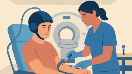
The operational flow starts in the MRI suite. Clinicians map peri-resection margins and infiltrative zones, define a treatment envelope, and plan sonication paths that respect anatomy and critical structures. During therapy, MRI supports both navigation and monitoring, offering thermal maps when needed and nonthermal readouts that confirm target coverage and procedural safety.
Microbubbles do the heavy lifting at the microscopic level. These gas-filled contrast agents, infused intravenously, oscillate under low-intensity ultrasound. The gentle, stable cavitation they create exerts mechanical stress on endothelial tight junctions, loosening them enough to let molecules pass for minutes to hours. Crucially, this is reversible. As the acoustic field turns off and bubbles clear, the junctions re-seal, restoring the barrier and limiting off-target exposure.
Safety is engineered into the protocol. Acoustic pressure is titrated to stay within non-destructive ranges, while real-time cavitation monitoring guards against inertial events that could bruise vessels or tissue. Dose control extends beyond sound energy to microbubble quantity and timing, and to how long the BBB-opening window stays active. This feedback stack aims for a repeatable, clinic-friendly procedure rather than a one-off heroics session.
Performance and Evidence From the Clinic
Evidence moved from promise to signal in a multicenter, open-label, phase 1/2 comparative trial led by a team spanning several North American institutions. Thirty-four patients with glioblastoma received BBB opening by FUS immediately before temozolomide during adjuvant cycles. Their outcomes were compared against 185 rigorously matched external controls treated with temozolomide alone after standard surgery and chemoradiation.
The differences were notable. Median progression-free survival stretched to roughly 14 months with FUS, compared with about 8 months in matched controls. Median overall survival exceeded 30 months in the FUS cohort, versus 19 months for controls. While matching cannot erase every bias that randomization handles, the magnitude and direction of benefit—paired with procedural feasibility over repeated monthly sessions—indicated that the approach did more than open a barrier; it shifted the clinical trajectory.
An additional insight came from blood. By sampling shortly after BBB opening, investigators detected tumor-derived signals that tracked with both progression-free and overall survival. That alignment suggests a workable path for liquid biopsy in a space where plasma-based assays have struggled. It is early, and assay standardization will matter, but the door appears open for pairing pharmacologic intensification with better surveillance.
Use Cases and Real-World Readiness
The most immediate fit is maintenance therapy for newly diagnosed disease. After recovery from chemoradiation, patients cycle through temozolomide; adding FUS before dosing targets the margins where recurrence incubates. The cadence—roughly monthly sessions for up to six cycles—matches existing workflows, which should ease adoption at centers with MRI capacity and trained staff.
Recurrent disease presents a separate opportunity. FUS could escort alternative chemotherapies, targeted small molecules, antibodies, or even viral vectors past the BBB. Agents abandoned for poor brain penetration might earn a second look under this delivery paradigm. Each combination will need careful pharmacokinetics and safety analysis, but the concept supports a portfolio strategy rather than a one-drug bet.
Liquid biopsy-enabled monitoring may change follow-up habits. Post-FUS blood draws that briefly enrich circulating tumor DNA and proteins could offer earlier signals of response or impending progression, complementing MRI and neurologic exams. With defined sampling windows and validated thresholds, clinicians could adapt therapy faster, potentially sparing patients from ineffective cycles.
Limitations, Risks, and What to Watch
Nonrandomized comparisons can be misleading, even with tight matching. Imbalances in molecular features such as MGMT promoter methylation or IDH status, differences in extent of resection, age, performance status, and institutional follow-up practices can tilt results. The interventional arm’s size was modest, which amplifies variance and narrows the margin for error. Randomized, stratified trials with blinded assessment remain the litmus test.
Safety also warrants ongoing scrutiny. The procedure’s risk profile centers on edema, petechial hemorrhage, or neurological symptoms from off-target or excessive cavitation. So far, events have aligned with expectations from preclinical and early clinical literature, and repeat sessions proved deliverable. However, expanding to broader populations and community settings will require standardized sonication parameters, microbubble dosing, MRI sequences, and stop rules that travel well across sites.
Finally, today’s protocols infer intraparenchymal exposure from the act of opening, not from per-patient drug level measurements. Direct pharmacokinetic confirmation inside the treated brain would sharpen dose optimization and could reveal interpatient differences in barrier dynamics. That knowledge would feed directly into more intelligent, patient-specific planning.
Market Dynamics and Ecosystem Momentum
The broader ecosystem appears readying for scale. Multiple centers now operate MRI-guided FUS platforms with shared training pathways, enabling multisite studies and, increasingly, routine care pilots. This platform plays well with oncology’s push toward drug repurposing: improving delivery for existing agents may unlock latent efficacy without wholesale changes in systemic dosing.
There is also a strategic upside for drug developers. A reliable BBB-opening companion could rescue candidates shelved for inadequate brain exposure and expand label opportunities for approved therapies. Payers and health systems will ask hard questions about cost, throughput, and durability of benefit, but a survival signal of the size reported—if confirmed—tends to move reimbursement discussions from “if” to “how.”
What Should Happen Next
The next phase is largely executional. Randomized, adequately powered trials should stratify by molecular markers, predefine imaging and blood-based endpoints, and include independent oversight. Pharmacokinetic and pharmacodynamic studies need to quantify how much drug enters, how long it stays, and how that relates to outcomes. Liquid biopsy workflows should be standardized—timing, assays, thresholds—so that signals are comparable across centers and actionable in clinic.
Technology will evolve in parallel. Expect more sophisticated feedback control, smarter patient selection based on anatomy and biomarker profiles, and AI-assisted targeting that reconciles tumor biology with acoustic constraints. With those tools, clinicians could tune not just where and how long to open the barrier, but also which drug, which dose, and which cycle offers the best trade-off for a given patient.
Verdict
MRI-guided focused ultrasound for transient BBB opening read as an inflection point for glioblastoma maintenance therapy rather than a niche maneuver. It delivered a credible survival signal when paired with temozolomide, stayed within manageable safety bounds, and unlocked a plausible route for blood-based monitoring in a disease long starved of it. The path forward depended on randomized validation, assay harmonization, and cost–workflow alignment, but the technology had crossed from intriguing to consequential and offered a practical, scalable way to push brain cancer care toward more targeted delivery and more responsive monitoring.