
Artificial intelligence (AI) is revolutionizing various sectors, and medicine is no exception. Researchers at UC San Francisco (UCSF) are at the forefront of integrating AI and machine learning with medical imaging to create human-centered AI solutions. These innovations aim to tackle some of the most significant challenges in medicine, including early illness detection, enhancing image quality for better diagnoses, non-invasive heart disease diagnosis, and monitoring the progression of neurodegenerative diseases. This article explores how AI is poised to transform these critical areas in medicine.
Early Illness Detection
AI in Pneumothorax Detection
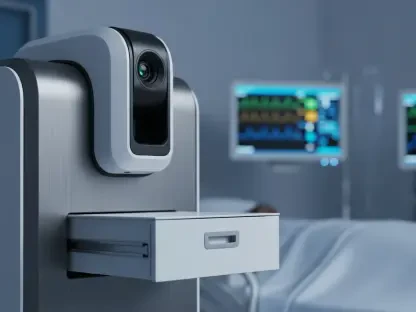
Diagnosing pneumothorax, a condition where the lung collapses, can be challenging due to its similarity to other conditions in symptoms and x-ray appearances. Radiologists, who interpret hundreds of images daily, might miss subtle signs of a collapsed lung. To address this, UCSF researchers developed the first AI bedside program to flag potential cases of pneumothorax for radiologists. Licensed by the FDA in 2019, this tool is now implemented in thousands of GE Healthcare machines worldwide. It works with portable X-ray machines, allowing doctors to use it directly at a patient’s bedside without significant infrastructure investments.
Traditional diagnosis methods for pneumothorax rely heavily on the expertise of radiologists to notice slight abnormalities in chest X-rays. However, the high volume of images they must analyze often results in missed diagnoses. Recognizing this issue, UCSF developed a technology that assists radiologists by flagging potential pneumothorax cases through AI. This groundbreaking tool leverages machine learning algorithms trained to identify the telltale signs of lung collapse from thousands of chest X-rays, thus enhancing the diagnostic process. Implementing this AI tool at the bedside accelerates diagnosis and improves patient outcomes by providing immediate insights that can spur prompt medical intervention.
Training the AI Tool
The AI tool was trained using a comprehensive database of thousands of anonymous chest X-rays, some showing collapsed lungs and others not. This training enabled the AI to recognize and flag potential cases accurately. By delivering quicker diagnoses and serving as an additional safety check, this development enhances patient care significantly.
AI models require vast amounts of data to become proficient, and the UCSF researchers compiled an extensive dataset of X-rays to develop their pneumothorax detection tool. The dataset included diverse cases where the lung collapse was present and absent, allowing the AI to learn the nuanced differences. Each image was meticulously labeled, providing the machine learning algorithms with the necessary information to distinguish healthy lungs from those exhibiting pneumothorax. Through repeated iterations, the AI improved its accuracy, learning to identify even the most subtle manifestations of the condition. This rigorous training process ensured the AI could function reliably in clinical settings, aiding radiologists by minimizing the chances of oversight and improving patient safety across healthcare facilities.
Enhancing Image Quality for Better Diagnoses
Improving MRI Resolution
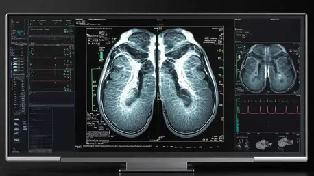
Standard MRI systems in the U.S. often operate at lower resolutions, which may miss crucial signs of conditions like multiple sclerosis and traumatic brain injuries. While higher resolution 7T MRI machines exist, their high cost limits their availability. UCSF researchers, led by Reza Abbasi-Asl, Ph.D., employed AI to enhance the resolution of standard MRIs, particularly for traumatic brain injury cases. This AI application made lower-resolution 3T MRI images comparable to high-resolution 7T images, outperforming other AI-enhanced MRI techniques.
Low-resolution MRI images often fail to capture detailed information necessary for an accurate diagnosis, especially in complex conditions such as multiple sclerosis and traumatic brain injuries. Recognizing the limitations of standard MRI machines, UCSF’s research team leveraged AI to bridge this gap. The AI system they developed enhances 3T MRI images, effectively bringing their quality closer to the highly coveted 7T scans. This technological leap means that high-resolution diagnostic imaging is no longer confined to facilities with expensive equipment. Patients everywhere benefit from clearer, more detailed scans, which translate into more precise diagnoses and timely treatments. Moreover, the ability to upscale lower-resolution images helps clinicians detect issues that might otherwise remain hidden, improving overall healthcare quality.
Machine Learning Models
The AI technique involves machine learning models that connect data patterns within paired MRI images of traumatic brain injuries. These pairs include a low-resolution 3T image and a high-resolution 7T counterpart. By constructing these models, researchers identified patterns and features challenging to detect in lower resolution images, enhancing image quality by focusing on specific details while minimizing noise. This advancement makes high-quality medical imaging more accessible without the need for expensive equipment.
By training machine learning models on paired datasets of 3T and 7T MRI images, researchers managed to teach AI to enhance image quality skillfully. The models learned to recognize specific features in low-resolution images by comparing them with their high-resolution counterparts. Through this process, AI became adept at amplifying details and reducing noise in 3T scans, which significantly improved their utility in clinical practice. This method holds potential beyond traumatic brain injury diagnosis; it could revolutionize how other conditions are identified, monitored, and managed by democratizing access to superior imaging technology. In a medical landscape constrained by equipment costs, AI-driven enhancements offer a cost-effective alternative ensuring accurate and comprehensive diagnostic capabilities.
Non-Invasive Heart Disease Diagnosis
AI in Coronary Artery Disease Diagnosis
Coronary artery disease, a leading cause of adult deaths globally, results from fatty deposits in the arteries that restrict blood flow to the heart. Physicians typically use coronary angiograms to visualize blood flow, but further invasive testing may be necessary to assess the heart’s left ventricle, posing risks to the kidneys. UCSF cardiologist Geoff Tison, M.D., MPH, and his team developed an AI model named CathEF to evaluate the heart’s pumping function by analyzing standard angiogram videos, eliminating the need for further invasive procedures.
Traditional coronary artery disease diagnosis often involves invasive techniques that carry inherent risks and discomfort for patients. Coronary angiograms, while effective, may require additional testing using contrast dye to assess heart function comprehensively. This extra dye can strain the kidneys, posing increased risks, especially in patients with preexisting conditions. Recognizing the need for a safer method, Dr. Geoff Tison and his team created CathEF, an AI-driven model that gauges heart performance using standard angiogram footage. CathEF leverages cutting-edge machine learning algorithms to analyze angiogram videos and generate accurate assessments of coronary artery function, potentially reducing the reliance on riskier, more invasive procedures and enhancing overall patient safety.
Deep Neural Networks
Deep neural networks, capable of learning complex data patterns in images and videos, were trained on anonymized angiogram videos. These networks accurately predicted the left ventricle’s pumping efficacy compared to ultrasound measurements, with consistent results in tests conducted outside the lab. CathEF represents a novel approach that uses routine angiogram data to provide additional vital information without requiring extra procedures, enhancing patient care.
The development of CathEF hinged on the sophisticated capabilities of deep neural networks, which excel at deciphering intricate patterns in visual data. Researchers trained these networks using anonymized angiogram videos to predict the heart’s pumping function, cross-verifying results with traditional ultrasound measurements. The AI’s ability to match and sometimes exceed the accuracy of conventional methods in clinical settings, including external tests, underscores its reliability. This innovative use of routine angiogram data not only simplifies the diagnostic process but also preserves patient well-being by reducing the need for invasive follow-up procedures. As AI technologies like CathEF become more integrated into medical practice, they promise to redefine cardiac care through enhanced, non-invasive diagnostics.
Monitoring Parkinson’s Disease Progression with Mobile Technology
AI in Neurodegenerative Disease Monitoring
Parkinson’s Disease, a neurodegenerative disorder affecting movement, requires accurate tracking of disease progression for optimal treatment. Physicians currently rely on intermittent patient visits and self-reported symptoms, which can miss subtle changes. UCSF researchers, including Simon Little, MBBS, Ph.D., and Reza Abbasi-Asl, Ph.D., developed machine learning techniques to monitor Parkinson’s Disease using smartphones and digital cameras. By capturing changes in gait and hand movements through recorded videos, the AI system can track disease progression and provide more precise data for tailored treatments.
Currently, the standard practice for monitoring Parkinson’s Disease involves sporadic clinical visits and subjective patient reports, which may not reflect the true extent of the disease’s progression. AI’s introduction into this sphere has the potential to revolutionize monitoring techniques by providing continuous and objective data. The AI system developed by UCSF researchers can analyze videos recorded via common digital devices to observe and quantify changes in patients’ movements, such as walking speed and hand mobility. This method allows for more consistent and detailed tracking, offering physicians a comprehensive view of their patients’ condition over time. By making precise adjustments to treatments based on accurate data, clinicians can significantly enhance the management and quality of life for those living with Parkinson’s Disease.
Machine Learning Programs
The research involved recruiting volunteers from the UCSF Movement Disorder and Neuromodulation Center. Participants were filmed performing common clinical examinations like walking and finger tapping. Machine learning programs processed these videos, isolating clinically relevant features such as finger tap speed that might indicate the disease’s severity. This digital transformation marks a departure from traditional, more subjective patient assessments, pointing towards a future where AI-enhanced monitoring becomes common practice in clinical settings.
In clinical trials, researchers captured videos of patients executing standard movement tests, which were then processed by sophisticated machine learning programs. These programs isolated critical features such as gait stability and finger-tapping speed, correlating them with Parkinson’s Disease severity. This approach not only offers a more accurate analysis than subjective reports but also equips clinicians with quantifiable data to refine treatment regimens. The shift towards AI-driven monitoring heralds a future where continuous, precise tracking of neurodegenerative diseases is feasible, enhancing both prognosis and patient care. As this technology evolves, its applicability may extend to other neurodegenerative disorders, promoting broader improvements in neurological healthcare.
Overarching Trends and Consensus Viewpoints
AI as a Supplemental Tool
A clear theme throughout these applications is AI’s ability to augment medical diagnostics and treatment plans. AI consistently enhances existing medical practices by providing more precise, efficient, and non-invasive methods for spotting diseases, diagnosing conditions, and tracking progress. This not only helps in delivering timely and accurate medical care but also in reducing the strain on healthcare resources and personnel.
The evident trend is AI serving as an adjunct to, rather than a replacement for, human medical expertise. AI systems enhance the precision and speed of diagnostics, enabling earlier detection of diseases and more tailored treatment plans. These intelligent tools support medical professionals by taking over repetitive tasks, freeing them to focus on complex decision-making and patient care. The integration of AI technologies in medical practice represents a harmonization of cutting-edge innovation with human intuition, aiming to deliver exceptional patient outcomes.
Conclusion
Artificial intelligence (AI) is making a significant impact across multiple sectors, and the field of medicine is no exception. At UC San Francisco (UCSF), researchers are leading the charge by combining AI and machine learning technologies with medical imaging. These advancements are geared toward creating human-centered AI solutions to address some of the most pressing challenges in healthcare. Key areas of focus include the early detection of illnesses, improving image quality for more accurate diagnoses, non-invasive procedures for diagnosing heart disease, and keeping track of the progression of neurodegenerative diseases.
One of the primary goals is to harness AI to detect diseases at an early stage when they are often more treatable. Enhanced image quality achieved through AI-powered tools enables doctors to make more accurate and timely diagnoses. In terms of heart disease, non-invasive methods driven by AI can spare patients from more invasive, riskier procedures, providing quick and precise assessments. Additionally, AI is proving invaluable in monitoring neurodegenerative conditions, offering new ways to track disease progression and potentially improve patient outcomes.
This article delves into the transformative potential of AI in these critical areas of medicine, illustrating how it can improve patient care and operational efficiency within the healthcare system.