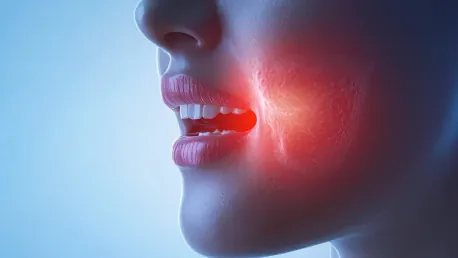

The transformative role of artificial intelligence (AI) and deep learning (DL) in the realm of medical imaging is ushering in a new era for the diagnosis and prognosis of oral potentially malignant disorders (OPMDs). These technologies are spearheading advancements in early detection and risk assessment for pre-cancerous lesions that can potentially progress to oral cancer. This innovative approach not only promises to enhance diagnostic accuracy but also to facilitate early intervention, crucial for improving patient outcomes.
The Role of AI in Medical Imaging
Clinical Photographic Images
AI applications, particularly convolutional neural networks (CNNs) such as DenseNet-169, ResNet-101, and EfficientNet-b4, have been employed to analyze clinical photographic images of oral lesions. These advanced CNN models have demonstrated an impressive ability to distinguish OPMDs from benign lesions and oral cancer. Studies reveal that the diagnostic sensitivity and specificity of these models often rival those of expert clinicians, marking a significant leap in the field. This high diagnostic accuracy, combined with the integration of smartphone-based imaging, presents a considerable advantage, especially in resource-limited settings where access to specialized healthcare services is restricted.
The adaptation of AI for clinical use not only enhances precision but also democratizes access to quality healthcare by reducing dependence on local expertise. Communities with limited medical infrastructure can particularly benefit from these advancements. As these technologies evolve, they hold the potential to make sophisticated diagnostic tools available globally, providing equitable healthcare opportunities and enabling early interventions that could substantially lower the morbidity and mortality associated with late-stage oral cancer diagnoses.
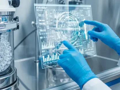
Autofluorescence Imaging
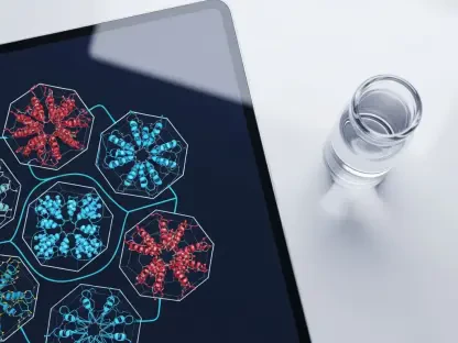
Another significant stride in AI-enhanced medical imaging is its application in autofluorescence imaging, an innovative technique that emphasizes biochemical changes within oral tissues. This method is augmented by AI to facilitate differentiation among normal mucosa, OPMDs, and malignant lesions with much-improved accuracy. Deep learning models trained on autofluorescence spectra are becoming integral in identifying subtle changes, thereby providing a valuable tool for early and precise detection of potentially malignant conditions.
These AI-supported enhancements not only improve diagnostic accuracy but also bridge critical gaps in the traditional assessment methods, which often rely heavily on subjective interpretation. By offering a more objective analysis, AI-enhanced autofluorescence imaging reduces diagnostic errors and ensures a higher standard of patient care. Furthermore, as technology integrates more deeply into clinical practices, it opens avenues for standardized diagnostic protocols that could harmonize the quality of care worldwide, particularly in regions lacking specialized medical infrastructure.
Noninvasive Diagnostic Techniques
Exfoliative Cytology
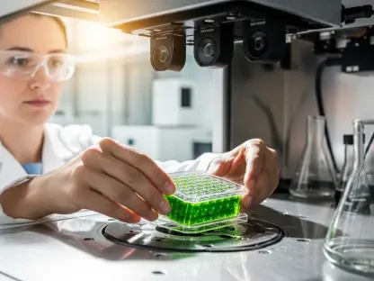
Exfoliative cytology, recognized for its noninvasive and cost-effective nature, involves the analysis of exfoliated cells from the oral mucosa. The introduction of AI into this domain has significantly enhanced the diagnostic accuracy of the method. CNN-based models have shown high sensitivity and specificity in detecting cytological changes that may indicate malignant transformation. This leap in technological integration means that noninvasive techniques are now nearly as reliable as more invasive methods, offering a patient-friendly alternative that maintains diagnostic rigor.
The advantages of AI-enhanced exfoliative cytology extend beyond comfort and cost-effectiveness; they also include streamlined workflows and faster diagnostic turnaround times. The integration of these AI-driven methods into routine clinical practice can alleviate the burden on histopathological analysis, allowing for quicker and more accessible preliminary screenings. This, in turn, facilitates prompt clinical decision-making, ultimately fostering better patient outcomes through earlier detection and intervention.
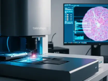
Optical Coherence Tomography (OCT)
Optical coherence tomography (OCT) stands as a high-resolution imaging modality adept at providing real-time images of oral tissues. AI models trained to analyze these OCT images have yielded diagnostic accuracies on par with those of seasoned pathologists. The ability to detect dysplastic and malignant changes at an early stage makes OCT a valuable tool in the clinician’s arsenal, particularly when enhanced with AI’s analytical prowess.
The amalgamation of AI with OCT extends beyond achieving comparable diagnostic accuracy with human expertise; it also introduces speed and consistency to the diagnostic process. This is achieved by mitigating the variability inherent in human interpretation and ensuring that significant pathological features are consistently identified and acted upon. Consequently, AI integrated with OCT paves the way for more standardized and reliable diagnostic practices, which are essential for the early detection and treatment of oral cancers.
Histopathological Analysis and Prediction Models
Histopathological Analysis
Despite the advent of various diagnostic technologies, pathological examination remains the gold standard for diagnosing OPMDs. However, deep learning algorithms offer a transformative approach by automating the detection of dysplastic features within histopathological images. Notably, models like Mask R-CNN have proven highly effective in identifying nuclear changes that indicate malignant potential, thereby reducing the variability associated with human interpretation.
This shift towards automation in histopathological analysis not only mitigates subjective variability but also significantly enhances throughput by rapidly processing and analyzing large volumes of data. Clinicians can thus focus on treatment decisions backed by robust, AI-driven insights. As the technology matures, AI-assisted histopathology is poised to become a cornerstone of diagnostic medicine, offering unparalleled precision and efficiency in identifying pathological changes.
Predicting Malignant Transformation
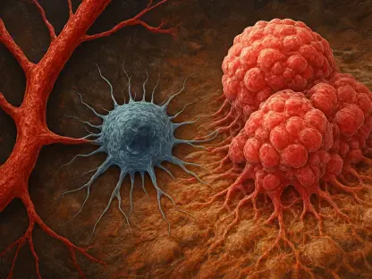
Beyond diagnosis, AI models are increasingly employed to predict the risk of OPMDs evolving into malignancies. Machine learning techniques, including random forest classifiers and survival models like DeepSurv, integrate various data types—clinical, histopathological, and imaging—to generate individualized risk assessments. These models provide clinicians with indispensable tools for personalized patient management, enabling tailored intervention strategies based on each patient’s unique risk profile.
The use of AI to predict malignant transformation embodies a shift towards proactive healthcare, where prevention and early intervention supersede reactive treatments. By anticipating the progression of OPMDs, healthcare providers can implement targeted surveillance and intervention measures, potentially thwarting the development of full-blown oral cancers. This proactive approach not only enhances patient outcomes but also optimizes resource allocation, focusing efforts where they are most needed.
Challenges and Future Directions
Data and Algorithmic Challenges
Despite the promising advancements in AI and deep learning, several challenges remain in seamlessly integrating these technologies into the diagnosis and prognosis of OPMDs. One of the foremost challenges is the need for large, standardized image datasets to train these models effectively. The variability in image quality across different sources and the inherent algorithmic limitations, such as overfitting and interpretability issues, also pose significant hurdles. These challenges necessitate concerted efforts in data collection, quality control, and algorithm refinement to ensure the reliability and robustness of AI applications in clinical practice.
Addressing these challenges requires a multidisciplinary approach, combining expertise in computer science, medical imaging, and clinical practice. Collaborative efforts to create comprehensive and standardized image repositories, along with advanced algorithmic techniques to enhance model interpretability and generalizability, will be crucial in overcoming these obstacles. Moreover, ongoing validation and real-world testing are essential to establish the clinical efficacy of AI-driven diagnostics, ultimately paving the way for broader adoption and integration into everyday practice.
Future Research Directions
The transformative impact of AI and DL on medical imaging is marking a new era in diagnosing and predicting oral potentially malignant disorders (OPMDs). These cutting-edge technologies are at the forefront of advancements in early detection and risk assessment of pre-cancerous lesions, which hold the potential to develop into oral cancer. This innovative method not only aims to enhance diagnostic precision but also supports early intervention, a factor that is vital for improving patient outcomes. By enabling doctors to identify these high-risk lesions early on, AI and DL are helping to catch and treat conditions that could otherwise progress to more severe stages, ultimately saving lives. Additionally, this technological leap is making it easier for healthcare professionals to provide personalized treatment plans based on individual risk profiles. As AI and DL continue to evolve, their role in medical imaging will likely expand, potentially leading to further improvements in patient care and management of OPMDs.