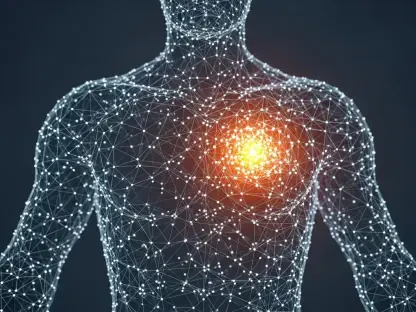

The transition from manual microscopy to digital oncology is no longer a futuristic concept but a present-day reality reshaping how patients experience cancer treatment from diagnosis to recovery. For over a century, pathologists have peered through lenses at slivers of tissue, but the integration of high-resolution scanning and artificial intelligence has effectively turned these biological specimens into high-value data for medical researchers. This metamorphosis is not merely a change in tools; it represents a paradigm shift toward computational pathology, where every pixel of a digital slide contains hidden biomarkers that can predict treatment responses or identify genetic mutations. As clinical settings across the globe move toward a digital-first approach in 2026, the traditional glass slide is becoming a digital artifact, enabling a faster, more accurate, and highly collaborative environment that promises to solve the long-standing bottlenecks of manual diagnostic reporting and subjective grading systems.
The Rise of Foundation Models and Scalable Intelligence
Modernizing Model Training: The Shift to Self-Supervision
Early attempts at implementing machine learning in oncology were often stifled by the grueling nature of supervised learning, which required highly trained pathologists to manually label thousands of individual cell structures. This “artisanal” methodology was not only time-consuming but also introduced human bias into the algorithmic baseline, making it difficult to produce tools for rarer forms of cancer where data sets were limited. However, the emergence of Foundation Models (FMs) has radically altered this trajectory by utilizing self-supervised learning techniques that allow algorithms to analyze millions of whole-slide images without the need for manual human annotation. By processing vast quantities of unlabelled data from diverse clinical archives, these models develop a foundational understanding of cellular architecture and tissue morphology, creating a numerical language that represents the biological essence of the sample. This development allows researchers to build highly accurate diagnostic tools for a wide array of cancers in a fraction of the time it previously took, effectively breaking the data bottleneck that once hindered progress in specialized oncology fields.
Building on this newfound efficiency, these advanced models act as a versatile computational backbone that can be rapidly adapted for specific clinical purposes with minimal additional data. Instead of starting from scratch for every new type of tumor, developers now use these pre-trained Foundation Models and fine-tune them for niche applications, such as identifying rare sarcomas or predicting immunotherapy responses in lung cancer. This modular approach to artificial intelligence development ensures that the technology can scale across the entire spectrum of oncology, providing a level of diagnostic support that was previously reserved only for the most common diseases. Furthermore, the transition to self-supervised learning significantly reduces the logistical burden on pathology departments, as they no longer need to dedicate hundreds of hours to labeling slides for machine training purposes. As a result, the cycle of innovation in cancer care has accelerated, leading to more robust and reliable tools that can be deployed into active clinical workflows with higher confidence than ever before.
Generalizability: Creating Reproducible Standards Across Platforms
One of the most persistent challenges in digital pathology has been the high degree of variability between different tissue staining methods and slide scanning hardware used across various hospitals. Historically, an AI model trained on slides from one institution might fail when applied to slides from another due to subtle differences in color, contrast, or image resolution, which limited the widespread adoption of these tools. To combat this, modern Foundation Models are trained on massive, heterogeneous datasets that encompass a wide variety of staining techniques, scanner brands, and anatomical sites, fostering a level of generalizability that was once considered unattainable. By exposing the AI to this diversity during its initial training phase, the resulting models become significantly more resilient to the noise of real-world clinical environments, ensuring consistent performance regardless of where the tissue sample was originally processed. This robustness is critical for establishing reproducible diagnostic processes, moving the field away from the subjective nature of manual microscopy and toward a standardized, quantitative science that yields the same results in any laboratory.
This shift toward reproducibility is not just a technical victory but a fundamental improvement in the reliability of patient care across different geographic regions. When diagnostic tools are able to maintain their accuracy across disparate platforms, it becomes possible to establish a unified standard of care that minimizes the risk of misdiagnosis or grading errors. Researchers are now able to develop high-quality models for rare pathological problems with the certainty that these tools will perform as expected when deployed in community hospitals or rural clinics. This maturation of technology has moved the industry from craft processes, which were highly dependent on specific localized conditions, to a system of reproducible processes that can be verified and validated on a global scale. In 2026, the focus has shifted from merely proving that AI can work to ensuring that it works consistently for every patient, regardless of the technological infrastructure of the treating facility, thereby laying the groundwork for a more equitable and scientifically rigorous approach to oncology.
Multimodal Integration and Enhanced Interaction
Breaking Data Silos: The Power of Multimodal Frameworks
The next frontier in the evolution of oncology involves the integration of disparate data types into a unified, multimodal artificial intelligence framework that provides a comprehensive view of the patient. Historically, the fields of pathology, genomics, radiology, and clinical history existed in isolated silos, requiring physicians to manually piece together information from multiple reports to make a single treatment decision. Modern AI systems are breaking these barriers by identifying latent patterns that bridge these different disciplines, such as subtle morphological signals within a tissue slide that correlate strongly with specific genetic mutations. By combining the visual data from pathology slides with complex genomic sequencing and radiomic imaging, these algorithms can detect nuances in the disease that are completely invisible to the human eye. This holistic approach allows for a level of personalized medicine that was previously impossible, as the AI can predict how a specific tumor will respond to a particular drug by analyzing the interplay between the patient’s genetic profile and the physical structure of the cancerous cells.
Furthermore, the implementation of multimodal frameworks is revolutionizing the way clinical trials are conducted and how patient outcomes are monitored over time. By analyzing thousands of data points simultaneously, these systems can identify patients who are most likely to benefit from experimental therapies, thereby increasing the success rate of drug development. This capability also extends to real-time clinical monitoring, where AI can track the evolution of a tumor through successive imaging and biopsy samples to detect signs of treatment resistance at the earliest possible stage. The synergy between different data streams ensures that no piece of information is overlooked, providing oncologists with a multidimensional map of the disease that guides more effective and targeted interventions. As these multimodal tools become more integrated into the daily operations of cancer centers in 2026, the focus is shifting toward proactive disease management, where data-driven insights allow clinicians to stay one step ahead of cancer’s ability to adapt and spread throughout the body.
Vision-Language Models: The New Diagnostic Dialogue
Innovative research is currently moving toward the development of Vision-Language Models (VLMs), which allow pathologists to interact with digital slides using natural language queries. Instead of relying on static reports or manual measurement tools, a pathologist can effectively hold a conversation with the diagnostic system, asking the model to find all areas of tumor-infiltrating lymphocytes or to describe the specific morphology of a suspicious region. This capability is made possible by encoding visual data and natural language into the same computational space, allowing the AI to understand the context of a pathologist’s request and provide immediate, relevant feedback. This interactive approach makes the analysis of complex digital slides as intuitive as a consultation with a human colleague, significantly reducing the time required to navigate through massive, high-resolution images. By serving as an intelligent interface between the doctor and the data, these models enhance the specialist’s ability to focus on the most critical aspects of a diagnosis while the machine handles the labor-intensive scanning and identification tasks.
This advancement in human-computer interaction represents a major step forward in making digital pathology more accessible and efficient for medical professionals of all experience levels. These tools act as a sophisticated assistant that can highlight areas of interest that might otherwise be missed during a standard visual review, providing a built-in second opinion that operates in real-time. The ability to use natural language to query complex datasets also facilitates better communication between members of a multidisciplinary care team, as the AI can generate summaries and visualizations that are easy for surgeons, oncologists, and radiologists to interpret. This shared understanding of the disease, facilitated by the bridge between visual evidence and clinical language, ensures that all members of the team are working from the same precise information. As these vision-language interfaces become more refined, they are transforming the diagnostic workflow into a collaborative process where the strengths of human intuition and algorithmic precision are seamlessly combined to provide the highest quality of patient care.
Institutional Adoption and the Philosophy of Augmentation
Digital Infrastructure: The Path to Total Adoption
The practical application of these advanced technologies is currently being realized through large-scale digitization efforts at leading medical institutions around the world. These facilities are undertaking the massive logistical challenge of scanning every glass slide produced in their labs and migrating their entire archives to cloud-based management systems. This transition is not just about replacing microscopes with computer monitors; it is about building a digital infrastructure that allows data to flow seamlessly between research laboratories and clinical departments. By centralizing this information, hospitals can utilize sophisticated AI models to organize, search, and analyze their entire pathology database, turning decades of physical records into a living library of oncology insights. This institutional commitment is essential for bridging the gap between experimental AI development and daily clinical practice, ensuring that the latest scientific breakthroughs are actually reaching the patients who need them.
As these pioneering centers move toward full digital adoption by 2027, they are creating a blueprint for the rest of the medical community to follow. The implementation of high-speed scanners and robust cloud storage solutions has become more cost-effective, allowing regional and community hospitals to begin their own digital transformations. This expansion of digital infrastructure is critical for the widespread deployment of AI tools, as the algorithms require a steady stream of high-quality digital images to function effectively. Furthermore, the shift to a digital-first workflow enables better data security and long-term preservation of patient samples, which can be re-analyzed years later as new diagnostic models are developed. This forward-looking approach ensures that the medical community is not only solving the problems of today but is also building a foundation for future discoveries that will continue to improve cancer survival rates. The total digitization of the pathology workflow is a necessary prerequisite for a future where every patient’s treatment is guided by the most advanced computational tools available.
Augmented Intelligence: Empowering the Human Expert
A fundamental pillar of the digital transition in oncology is the philosophy of augmented intelligence, which emphasizes the use of technology to enhance rather than replace the role of the medical professional. This distinction is vital for maintaining the highest ethical and clinical standards, as it ensures that the final diagnostic interpretation and treatment decisions remain the responsibility of a qualified physician. In this model, the AI handles the repetitive, granular, and cognitively taxing tasks that are prone to human fatigue, such as counting thousands of individual cells or meticulously measuring tumor margins across multiple slides. By automating these “low-level” tasks, the technology frees up the pathologist to focus on “high-level” clinical judgment, complex case interpretation, and meaningful communication with the patient’s care team. This synergy creates a more efficient and accurate diagnostic environment where the speed and pattern-recognition capabilities of the machine are balanced by the experience and nuanced understanding of the human expert.
The implementation of augmented intelligence also serves as a critical safety net, providing a reliable second opinion that can flag subtle abnormalities that might be overlooked during a manual review. For example, AI can be programmed to scan an entire slide for minute clusters of metastatic cancer cells that are only a few dozen microns in size, highlighting them for the pathologist’s immediate attention. This collaborative workflow reduces the likelihood of diagnostic errors and ensures that every patient receives a thorough and meticulous evaluation of their tissue samples. Moreover, as AI models become more proficient at providing data-driven insights, they act as an educational resource for medical residents and fellows, who can use the technology to validate their own observations and learn from the massive datasets used to train the algorithms. By framing the technology as a capable assistant rather than a replacement, the medical community has fostered a culture of innovation that prioritizes patient safety and clinical excellence above all else.
Democracy and Economics in Precision Oncology
Universal Expertise: Overcoming Geographic Barriers
One of the most profound and far-reaching impacts of digital pathology is its ability to democratize access to high-quality oncological expertise across the globe. Historically, the most specialized knowledge in rare or complex cancers has been concentrated in elite academic medical centers located in major metropolitan areas, leaving patients in rural or underserved regions at a disadvantage. However, once a pathology slide is digitized, it can be transmitted across the internet in seconds, allowing it to be analyzed by the same sophisticated AI models and remote specialists regardless of where the patient is physically located. This removes the geographic barriers to precision medicine, ensuring that a patient in a remote village or a small community hospital can receive the same level of diagnostic scrutiny as someone treated at a world-renowned cancer center. This shift toward a decentralized model of care is essential for reducing healthcare disparities and ensuring that the benefits of technological progress are shared equitably across all populations.
This democratization of care also facilitates global collaboration on rare diseases, as researchers from different continents can easily share and analyze digital samples to identify common patterns and potential treatments. In 2026, the ability to consult with international experts through digital pathology platforms has become a standard practice for managing difficult cases, leading to more accurate diagnoses and better-informed treatment plans. Furthermore, the use of AI as a diagnostic baseline provides a consistent standard of care in regions where there may be a shortage of trained pathologists, helping to fill the gap in medical resources. By exporting specialized diagnostic power through the cloud, the medical community is creating a universal network of expertise that protects patients from the limitations of their local healthcare infrastructure. This globalized approach to oncology not only saves lives but also accelerates the collective understanding of cancer by allowing for the study of a much more diverse and representative range of patient populations.
Economic Efficiency: Reducing the Cost of Precision Medicine
The widespread adoption of AI-driven pathology offers a significant economic advantage that could make precision oncology more sustainable and accessible for healthcare systems worldwide. Currently, comprehensive genetic testing and molecular sequencing can cost thousands of dollars per patient, often making these advanced diagnostics unaffordable for many individuals and public health programs. In contrast, a standard pathology slide is relatively inexpensive to produce and contains a wealth of untapped information that AI is now beginning to unlock. As algorithms become increasingly proficient at identifying genomic-level insights—such as specific mutations or biomarkers—directly from these low-cost digital images, the need for expensive secondary molecular testing may be significantly reduced. This ability to extract high-level data from standard clinical materials represents a major breakthrough in cost-efficient healthcare, allowing for the delivery of personalized treatment plans at a fraction of the traditional price.
Beyond the reduction in direct testing costs, the efficiency gains provided by AI also lead to broader economic benefits for the healthcare system by streamlining the diagnostic process and reducing the time to treatment. Faster and more accurate diagnoses mean that patients can begin the correct therapy sooner, which often leads to better outcomes and lower long-term care costs associated with advanced-stage disease. By automating the most time-consuming aspects of the pathology workflow, institutions can handle a higher volume of cases with the same number of staff, helping to alleviate the economic pressure caused by a growing global cancer burden. This shift toward a more efficient, data-driven diagnostic model ensures that resources are allocated where they are most effective, maximizing the impact of healthcare spending. Ultimately, the economic disruption caused by digital pathology is paving the way for a future where the most advanced oncology care is a financially viable option for all patients, regardless of their economic circumstances or the wealth of their nation.
Future Outlook for a Digital Ecosystem
Technological Maturation: Moving Toward Global Deployment
The consensus among medical experts in 2026 is that the total digitization of the oncology landscape is an inevitable and essential step toward a modern healthcare ecosystem. Over the past few years, the advent of faster, more affordable slide scanners and the expansion of high-speed satellite internet have brought digital pathology within reach of regional hospitals and developing nations, moving the technology far beyond its origins in elite research centers. There is a concerted effort within the industry to distill massive, complex AI models into smaller, more efficient versions that can be deployed on standard hospital servers without the need for specialized supercomputers. This push for deployability ensures that the benefits of AI are not limited by a facility’s computational power, allowing even smaller clinics to integrate sophisticated diagnostic tools into their daily routines. As these technologies continue to mature and become more user-friendly, the friction associated with adopting new digital workflows is rapidly disappearing.
The maturation of this technology also involves the establishment of clear regulatory frameworks and standardized protocols that ensure the safety and reliability of AI-driven diagnostics across different jurisdictions. International health organizations have worked to create guidelines for the validation of these tools, providing a roadmap for hospitals to implement AI with confidence. This regulatory clarity has encouraged more institutions to invest in digital infrastructure, leading to a surge in the availability of high-quality data for further research and development. As the global digital ecosystem expands, the collective intelligence of the medical community is growing at an exponential rate, with every new digital slide contributing to a more comprehensive understanding of cancer biology. This interconnected network of data and expertise is the foundation of a new era in medicine, where the limitations of individual human observation are surpassed by the collaborative power of a global digital community.
Conclusion: Building a Universal Standard of Oncology Care
The journey toward a fully digital pathology ecosystem reached a critical turning point as institutions successfully moved beyond the experimental phase and into widespread clinical deployment. By converting physical tissue samples into computable data, the medical community effectively unlocked a level of diagnostic precision that was previously considered unattainable within the constraints of manual microscopy. The groundwork for this transformation was laid through the development of robust Foundation Models, the integration of multimodal data streams, and a steadfast commitment to the philosophy of augmented intelligence. These advancements ensured that the transition to a digital workflow did not diminish the human element of medicine but instead empowered clinicians with the tools necessary to meet the challenges of an increasingly complex diagnostic landscape. The results of these efforts were seen in the significant improvements in diagnostic accuracy, the acceleration of drug development, and the expansion of high-quality care to previously underserved populations.
Looking back at the progress made by 2026, it became clear that the digitization of cancer care was the most significant move toward a universal standard of oncology in modern history. The industry successfully demonstrated that precision medicine could be both highly advanced and economically accessible by leveraging artificial intelligence to extract deep insights from standard clinical materials. This shift effectively broke the geographic and financial barriers that once limited the reach of the most effective cancer treatments, making the highest standards of care a basic right for patients everywhere. As the medical community continued to refine these tools and expand the global digital network, the focus remained on the actionable goal of ensuring that every diagnostic insight was used to its fullest potential. The era of the glass slide had served its purpose for over a century, but the transition to a digital-first world provided the necessary foundation for a future where cancer diagnosis is faster, more accurate, and entirely focused on the unique needs of the individual patient.