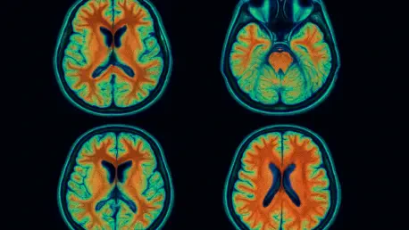

In the ever-evolving field of neuroscience, capturing the brain’s intricate workings in a natural, unaltered state has remained a formidable challenge for researchers striving to unlock its deepest secrets, while traditional imaging techniques, often reliant on anesthesia, introduce distortions that skew critical physiological processes. These processes, such as blood flow and neuronal activity, often result in data that may not fully represent real brain function. However, a groundbreaking advancement from The Hong Kong University of Science and Technology (HKUST), spearheaded by Professor Jianan Qu, has introduced a revolutionary high-speed imaging technology that captures high-resolution brain activity in awake mice. This innovation marks a significant departure from past limitations, offering a clearer, more accurate window into the brain’s natural operations. By eliminating the need for anesthesia, this method ensures that the observed activities reflect authentic physiological states, paving the way for more reliable research outcomes. The implications of such a breakthrough are vast, promising to reshape how brain functions and disorders are studied and understood in model organisms like mice, which are pivotal for human health research.
Revolutionizing Neuroscience with Awake Imaging
The ability to image the brain of awake mice without the confounding effects of anesthesia represents a monumental leap forward in neuroscience research. For years, anesthesia has been a necessary but problematic tool in brain studies, altering vital functions such as circulation and neuronal firing patterns, which in turn compromises the validity of experimental results. The technology developed by the HKUST team circumvents this issue entirely, allowing scientists to observe brain activity as it occurs naturally. This shift is particularly significant because mice serve as essential models for understanding human brain conditions, including neurodegenerative diseases and mental health disorders. With this new imaging approach, the data collected mirrors real-life scenarios more closely, enhancing the credibility of findings that could translate to human applications. The removal of anesthesia as a variable not only improves accuracy but also opens up new avenues for exploring how the brain responds to stimuli in an unmedicated state, setting a new standard for experimental design.
Beyond the immediate benefit of avoiding anesthesia, this technology offers a profound opportunity to delve into the brain’s dynamic processes with unprecedented precision. The high-speed imaging system ensures that even the subtlest activities within the brain are captured without distortion, providing a detailed view of interactions that were previously obscured. This capability is crucial for studying complex neurological phenomena, such as how different brain regions communicate during cognitive tasks or sensory processing. Furthermore, the method supports longitudinal studies where the same subject can be observed over time without the cumulative effects of repeated anesthesia, which often introduces health complications. Such advancements mean that researchers can now track the progression of brain-related conditions in a more naturalistic context, yielding insights that are more applicable to therapeutic development. This approach fundamentally changes the landscape of neuroscience, offering a tool that aligns experimental conditions with the brain’s inherent functionality.
Unveiling the Power of Multiplexing Digital Focus Sensing and Shaping (MD-FSS)
At the core of this transformative imaging breakthrough lies Multiplexing Digital Focus Sensing and Shaping (MD-FSS), a cutting-edge technique that redefines the speed and clarity of brain imaging. By employing multiple laser beams encoded at unique frequencies, MD-FSS rapidly measures the point spread function (PSF), a critical parameter for achieving sharp, high-resolution images. Astonishingly, this process is completed in under 0.1 seconds, a speed that outpaces previous methods by a factor of ten. This rapid data collection is essential when imaging awake mice, whose natural movements could otherwise blur results. The precision of MD-FSS ensures that every detail of brain activity is captured with clarity, even under dynamic conditions. This technological marvel not only enhances the quality of images but also sets a new benchmark for real-time observation, allowing researchers to study the brain as it actively processes information in a living subject.
The integration of MD-FSS with adaptive optics and three-photon microscopy further amplifies its impact by overcoming longstanding barriers in brain imaging. Traditional tools like MRI or EEG often fall short in providing the fine detail needed to visualize intricate brain structures, while the skull’s density obstructs light in conventional microscopy methods. This innovative combination, however, penetrates deeper into brain tissue and delivers exceptional image quality without the need for invasive procedures. The technology counters issues like light scattering, ensuring that researchers can explore the brain’s inner workings at a subcellular level. Such advancements are pivotal for neuroscience, as they enable a closer examination of critical components like neurons and glial cells in their natural environment. By providing a non-invasive means to achieve high-resolution imaging, MD-FSS stands as a testament to the potential of modern optical technologies to revolutionize how brain activity is studied and understood.
Overcoming Historical Challenges in Brain Imaging
Brain imaging has long been hindered by significant obstacles, such as the skull’s thickness, which impedes light penetration, and the physiological distortions introduced by anesthesia. Techniques like two-photon microscopy have struggled to maintain image quality due to these barriers, often resulting in blurred or incomplete data. The HKUST team’s pioneering approach, however, integrates adaptive optics with advanced three-photon microscopy to address these challenges directly. This synergy allows for deeper light penetration through the skull, capturing detailed images of brain structures that were previously inaccessible without invasive methods. By tackling such fundamental issues, this technology provides a clearer, more accurate representation of brain activity, enabling researchers to study intricate details that older methods could not reveal. This breakthrough is a critical step forward, bridging the gap between technological limitations and the need for precise neurological data.
Another persistent challenge in brain imaging has been motion blur, particularly when studying living, awake animals that move naturally during experiments. The high-speed capabilities of MD-FSS prove instrumental in this regard, ensuring that even the slightest movements do not compromise image quality. This rapid imaging process captures real-time brain activity with remarkable fidelity, allowing for the observation of dynamic processes as they unfold. Such precision is vital for understanding how the brain operates under normal conditions, without the interference of sedation or restraint. The ability to account for motion also enhances the reliability of data collected over extended periods, as it reduces artifacts that could skew results. This technological advancement marks a significant departure from past constraints, offering a robust solution for studying the brain in action and paving the way for more accurate interpretations of neurological functions.
Exploring New Frontiers in Neuroscience Research
The capacity to image awake mice at a subcellular level unlocks a treasure trove of information about brain function that was previously out of reach. This technology enables detailed tracking of immune cells like microglia, which play a key role in brain health, as well as monitoring blood flow dynamics in cerebral vessels. Additionally, it facilitates observation of neuronal behavior during sensory processing or cognitive tasks, shedding light on how different brain components interact in real time. These insights are invaluable for unraveling the complexities of brain networks and their responses to various stimuli. By providing a window into these intricate processes, the imaging method offers a deeper understanding of how the brain maintains balance and responds to challenges, which is essential for advancing knowledge in both basic and applied neuroscience. Such detailed data can inform models of brain function that are more aligned with natural physiological states.
The implications of this imaging technology extend far beyond basic research, significantly enhancing the study of neurological disorders. Conditions like Alzheimer’s, Huntington’s disease, and epilepsy can now be explored with greater accuracy, as the technology captures brain activity without the confounding effects of anesthesia. This clarity is crucial for identifying the mechanisms underlying these disorders and evaluating potential treatments. Furthermore, the method supports broader biomedical research, including investigations into cancer therapies and vaccine efficacy through mouse models. By offering a reliable platform for observing brain responses to experimental interventions, this technology aids in the development of targeted therapies that could improve outcomes for a range of conditions. The ability to study awake mice in such detail ensures that findings are more translatable to human contexts, strengthening the link between preclinical studies and clinical applications.
Future Horizons: Scaling Up for Greater Impact
Looking to the future, the scalability of MD-FSS holds immense promise for expanding the scope of brain imaging research. Currently designed to operate with eight laser beams for PSF measurement, the system has the potential to incorporate dozens or even hundreds of beams as light-control technologies advance. This expansion could enable coverage of larger brain regions and capture faster, more complex events, providing a comprehensive view of neural activity across extensive networks. Such scalability positions MD-FSS as a versatile platform for addressing some of the most pressing questions in neuroscience, from understanding learning and memory to mapping disease progression. The ability to adapt and enhance the system over time ensures that it remains at the forefront of technological innovation, ready to meet the evolving needs of researchers aiming to explore the brain’s vast intricacies.
The long-term impact of this imaging technology also lies in its potential to integrate with other functional assays, further enriching the data it can provide. As the system scales, it could be paired with techniques that measure biochemical changes or electrical activity, offering a multi-dimensional perspective on brain function. This holistic approach would be instrumental in studying rapid brain events and complex interactions that occur during normal and pathological states. The forward-looking design of MD-FSS suggests that it could redefine research methodologies, fostering collaborations across disciplines to tackle challenges in mental health and neurological disorders. By laying the groundwork for such advancements, this technology not only addresses current limitations but also anticipates future demands, ensuring that neuroscience continues to progress with tools that match the complexity of the brain itself.
Reflecting on a Transformative Milestone
Reflecting on the strides made by the HKUST research team, it becomes evident that their development of high-speed imaging technology marks a pivotal moment in neuroscience. The introduction of MD-FSS and its integration with adaptive optics and three-photon microscopy tackled longstanding barriers, delivering high-resolution insights into the brain activity of awake mice. This achievement eliminated the distortions of anesthesia, providing data that mirrored the brain’s natural state with remarkable accuracy. The detailed observations of neuronal dynamics, glial interactions, and vascular functions offered a clearer understanding of brain processes, setting a new benchmark for research precision. As this technology gains traction, it reshapes experimental approaches, ensuring that findings hold greater relevance for human health applications. Looking ahead, the focus should shift toward integrating this tool with emerging methodologies to deepen insights, while continued investment in scaling MD-FSS could unlock even broader brain regions for study, driving innovative solutions for neurological challenges.