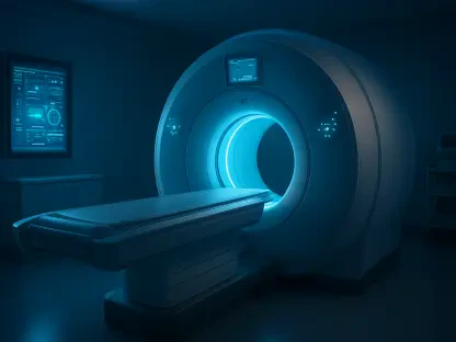

Magnetic resonance imaging’s multiplanar capabilities allow for the identification of tubular duplications that run parallel to the segment of the fetal bowel. This sophisticated imaging modality has fundamentally shifted the diagnostic landscape for clinicians managing rare congenital anomalies within the fetal abdomen. Enteric duplication cysts, or EDCs, represent a significant clinical puzzle because their appearance often mimics more common occurrences like ovarian cysts, mesenteric cysts, or meconium pseudocysts. These fluid-filled structures are lined with alimentary tract epithelium and can emerge anywhere from the oral cavity to the distal rectum. For decades, the difficulty in distinguishing these cysts from simple bowel loops led to delayed postnatal interventions. However, the integration of advanced fetal MRI protocols now provides the high-resolution detail needed to visualize the structural nuances of the gut wall. This clarity is paramount for early intervention planning and ensuring that neonates receive specialized care.
Evaluating Methodological Rigor and Regional Trends
Current diagnostic standards in 2026 rely heavily on comprehensive data sets that correlate prenatal imaging with definitive postnatal pathological findings. Recent longitudinal studies tracking fetal development from 2026 to 2028 have emphasized the importance of third-trimester scans, specifically between the twenty-sixth and thirty-ninth weeks of gestation. During this window, the anatomical maturation of the fetal gastrointestinal tract reaches a state where MRI can effectively differentiate between various tissue layers. This period is critical because the resolution of the image depends on the relative size of the abdominal structures compared to the surrounding amniotic fluid. Research involving pathologically confirmed cases has demonstrated a high degree of correlation between MRI signatures and the actual biological structure of these duplications. This evidence-based approach ensures that the diagnosis is not merely a clinical suspicion but a grounded medical finding that can guide subsequent surgical efforts.
Anatomical trends observed in recent clinical cohorts suggest a notable concentration of enteric duplication cysts in the ileocecal region, which comprises the distal portion of the small intestine. Statistical analyses indicate that over three-quarters of diagnosed cases are situated in this specific area, providing radiologists with a powerful diagnostic clue. From an embryological perspective, these malformations are thought to arise from errors occurring during the recanalization process of the primitive gut. Because the majority of these cysts are located on the mesenteric side of the ileum, their presence in this vicinity serves as a red flag for duplication rather than other generic cystic masses. Furthermore, the data continues to show a slight female predominance in these anomalies, aligning with historical patterns in gastrointestinal development. Recognizing these regional patterns allows medical teams to narrow their differential diagnosis quickly, focusing on the most likely anatomical culprits.
Morphological Variants and Structural Characteristics
The morphological diversity of enteric duplication cysts is typically categorized into two distinct forms: cystic and tubular. Cystic duplications are the most prevalent, appearing as discrete, spherical, or oval fluid-filled structures that remain separate from the primary intestinal lumen. In contrast, tubular duplications, which account for nearly twenty percent of cases, run parallel to the segment of the bowel and often share a more complex anatomical relationship with the parent organ. This distinction is far from academic; tubular forms are significantly more likely to communicate directly with the intestinal tract. Such communication poses unique postnatal risks, including the potential for internal bleeding if the lining contains heterotopic gastric mucosa, or the risk of acute inflammation and secondary obstruction. By identifying these shapes prenatally, radiologists provide surgeons with essential information regarding the potential for early-onset complications immediately following birth.
Beyond their basic shape, these anomalies are further evaluated based on their position relative to the intestinal wall, being classified as either extraluminal or intraluminal. Extraluminal cysts reside entirely outside the intestinal wall, while intraluminal variants develop within the muscular layers of the bowel itself. While many of these duplications are discovered as isolated findings, approximately fourteen percent of cases are accompanied by other structural malformations. This association necessitates a meticulous “top-to-bottom” survey of the entire fetus to ensure no other developmental disturbances are overlooked. Understanding whether a cyst is an isolated occurrence or part of a broader syndromic presentation is vital for long-term prognosis. This comprehensive diagnostic approach helps in establishing a more holistic view of the fetal health status, allowing for a multidisciplinary response that includes neonatologists, pediatric surgeons, and genetic counselors to manage the case effectively.
Decoding the Tissue Signatures of Enteric Cysts
A hallmark of fetal MRI is its ability to produce a refined signal profile that facilitates the differentiation of enteric duplication cysts from other abdominal pathologies. These lesions characteristically exhibit a homogeneous long T1 signal, which appears dark on the imaging screen, and a long T2 signal, which manifests as a bright, clear area. This specific signal combination is indicative of simple or near-simple fluid content, which is essential for ruling out masses that contain high concentrations of protein, fat, or calcification. For instance, meconium pseudocysts often display more heterogeneous signals due to the presence of debris or calcified elements, while simple enteric cysts remain clear and uniform. By utilizing these varying pulse sequences, clinicians can gain insight into the internal composition of the mass without invasive procedures. This level of tissue characterization is a primary reason why MRI has become the preferred secondary imaging tool following an initial ultrasound screening.
The most definitive characteristic that identifies an enteric duplication cyst on an MRI is the thickness and layered composition of its wall. Unlike mesenteric or ovarian cysts, which possess extremely thin serosal walls, an EDC features a robust wall that mimics the histological layers of the intestine, including the mucosa and the muscularis propria. On high-resolution scans, this “thick wall” sign is a reliable indicator that the mass is of enteric origin. Furthermore, these cysts almost always share a common wall with the adjacent bowel or appear directly continuous with it, emphasizing their mesenteric location. This intimate anatomical relationship is frequently cited as the “gold standard” for diagnosis, as it clearly distinguishes the duplication from other pelvic or abdominal structures that would be physically separate from the intestinal loops. Identifying this shared boundary provides the anatomical certainty required to confirm the diagnosis and prepare for the specific surgical needs of the newborn.
Strategic Planning and Postnatal Outcome Optimization
While ultrasound remains the ubiquitous first-line tool for prenatal screening, it is frequently hampered by factors such as maternal body habitus, low amniotic fluid levels, or the physical orientation of the fetus. MRI effectively circumvents these limitations by offering multiplanar imaging that is independent of the fetal position, providing a clear and unobstructed view of the abdominal anatomy. The superior soft-tissue contrast inherent in MRI allows for a precise measurement of the cyst’s dimensions and its proximity to major vascular structures. This level of detail is indispensable for the differential diagnosis process, enabling medical professionals to rule out mimics that might otherwise lead to incorrect surgical planning. By providing a three-dimensional understanding of the lesion’s location and its relationship to the surrounding organs, MRI ensures that the clinical team is not surprised by unexpected anatomical findings during postnatal intervention, thereby increasing the safety of the surgical procedure.
Effective prenatal diagnosis through MRI also serves as a critical foundation for parental counseling and the coordination of neonatal care. When an enteric duplication cyst was identified with high confidence, clinicians were able to provide families with a realistic and generally positive prognosis, as these lesions are typically benign and highly treatable through surgical resection. Knowing the exact location and classification of the cyst allowed for delivery to be scheduled at a facility equipped with specialized pediatric surgical teams ready for immediate evaluation. This proactive strategy minimized the risk of emergency interventions and ensured that the transition from delivery to surgery was seamless. The ability to distinguish between simple and complex duplications also helped in prioritizing surgical cases based on the risk of obstruction or hemorrhage. Ultimately, the precision of prenatal MRI transformed the management of these rare anomalies into a predictable and highly successful clinical pathway for both the infants and their families.
Integrating Advanced Imaging into Standard Care Protocols
The systematic implementation of fetal MRI protocols established a new benchmark for the accuracy of diagnosing enteric duplication cysts. By focusing on wall thickness, mesenteric attachment, and specific T2 signal characteristics, radiologists successfully narrowed the gap between imaging and pathology. This progress ensured that the identification of abdominal masses was no longer a matter of clinical guesswork but a data-driven process. Future protocols will likely incorporate AI-assisted image analysis to further refine the detection of these subtle tissue layers, potentially identifying anomalies even earlier in the second trimester. Clinicians should now view MRI not as an optional luxury, but as a necessary component of the diagnostic workup for any suspicious fetal abdominal cyst. As these imaging technologies continue to evolve, the emphasis must remain on the early and accurate classification of lesions to optimize postnatal outcomes. Continued collaboration between radiologists and pediatric surgeons will be essential.
Standardizing these imaging protocols across tertiary care centers ensures that every patient benefits from a high level of diagnostic accuracy regardless of geographic location. The transition from subjective interpretation to objective, signal-based diagnosis has already reduced the incidence of misdiagnosed ovarian cysts and improved surgical outcomes in the current 2026 medical landscape. Future efforts should prioritize the training of specialized radiologists who can navigate the complexities of fetal gut anatomy with precision. By fostering an environment where imaging data is seamlessly shared between specialists, the medical community can continue to build more robust predictive models for congenital anomalies. These actionable steps will bridge the gap between prenatal detection and long-term neonatal health, creating a more efficient healthcare journey for families facing these rare diagnoses. As the field moves forward, the commitment to refining these non-invasive techniques will remain the cornerstone of pediatric radiology.