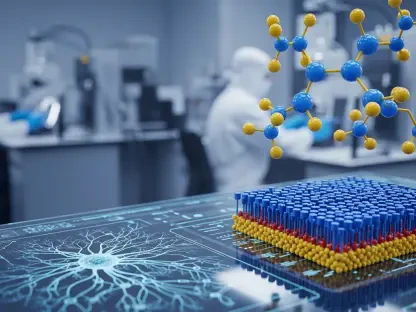

The advancements in artificial intelligence (AI) techniques, particularly in the analysis of medical imagery to enhance tumor detection in positron emission tomography (PET) and computed tomography (CT) scans, are set to revolutionize the field. These advancements are especially relevant due to the crucial role that imaging techniques play in cancer diagnosis and treatment. The integration of AI into these processes promises to enhance precision and efficiency, ultimately improving patient outcomes and possibly reshaping how medical professionals approach cancer detection.
The Role of PET and CT Scans in Oncology
PET and CT scans are indispensable tools in oncology, playing pivotal roles in diagnosing and managing various types of cancer. PET scans are instrumental in visualizing metabolic processes within the body, which is crucial for identifying malignant tumors. These tumors typically exhibit a higher metabolic rate than benign tissues, making them more detectable on PET scans. On the other hand, CT scans offer detailed images of the body’s anatomy, enabling the precise localization of tumors. The combination of these imaging techniques, when augmented with AI, particularly deep learning algorithms that utilize multi-layered artificial neural networks, shows significant potential for advancing the detection and analysis of tumors.
Incorporating AI into PET/CT imaging harnesses the power of vast datasets to recognize complex patterns, providing a more accurate and comprehensive understanding of tumor characteristics. Deep learning models can be trained to distinguish between malignant and benign tissues with a higher degree of precision than traditional methods. This technological convergence promises not only to improve detection rates but also to reduce false positives, thereby minimizing unnecessary treatments and patient anxiety. As these AI-driven methods continue to evolve, they are poised to become integral components in the field of medical imaging, offering a transformative approach to oncology diagnostics.
The Labor-Intensive Process of Tumor Identification
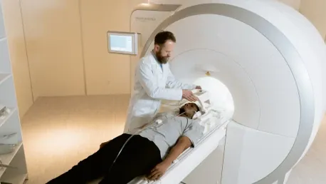
The current methods employed by doctors to identify and measure tumor lesions in PET and CT scans are notably labor-intensive. Physicians often have to manually mark 2D slice images to measure tumor size, a process that can become extremely time-consuming, particularly for patients with numerous lesions. This step is essential to forming a comprehensive picture of the cancer’s extent but is highly susceptible to human error and variability. The demand for precision in this manual process can be taxing on medical professionals, leading to variability in diagnoses and potentially impacting treatment outcomes for patients.
The introduction of AI into this domain offers a promising solution by automating the analysis of medical images. AI algorithms can be trained to perform the tedious tasks of highlighting and measuring tumor lesions with greater consistency and speed. Automation through AI could save a significant amount of time for radiologists, allowing them to focus on more complex aspects of patient care. Additionally, minimizing human error enhances the reliability of diagnoses, ensuring that critical decisions regarding treatment are based on accurate and consistent data. The shift from manual to automated processes heralds a new era of efficiency and precision in medical imaging, ultimately benefiting both healthcare providers and patients.
The Potential of AI in Enhancing Accuracy and Efficiency
Automation of medical image analysis through AI can lead to uniform and precise identification of lesions, potentially improving patient outcomes and streamlining therapeutic decisions. Professor Rainer Stiefelhagen, Head of the Computer Vision for Human-Computer Interaction Lab (cv:hci) at KIT, particularly emphasizes this potential. During the AutoPET competition, various teams were tasked with automatically segmenting tumor lesions in whole-body PET/CT images. The team of researchers from KIT, in collaboration with the Essen-based Institute for Artificial Intelligence in Medicine (IKIM), utilized deep learning methods to analyze a substantial annotated PET/CT dataset.
Their approach exemplifies how AI can enhance the efficiency of image analysis while maintaining or even improving accuracy. The deep learning models employed in the competition were designed to handle large, complex datasets, identifying patterns that may be imperceptible to the human eye. This capability is critical in detecting early-stage tumors and other subtle anomalies that might be missed during manual evaluations. The application of AI in this context goes beyond merely replicating human expertise; it augments it, providing medical professionals with powerful tools to deliver better care.
The Success of Ensemble Algorithms in Tumor Detection
The AutoPET competition results revealed a fascinating insight into the effectiveness of ensemble algorithms in tumor detection. An ensemble of top-rated algorithms outperformed individual algorithms by a considerable margin, both in terms of efficiency and accuracy. This ensemble method aggregates the strengths of multiple algorithms, resulting in more robust and reliable tumor detection. It highlights the importance of algorithmic design, where the interplay of various models and their unique advantages are integrated to produce superior outcomes, as opposed to relying on a single algorithmic approach.
This finding underscores the significance of collaborative and diversified approaches in AI development. By leveraging the distinct capabilities of various models, the ensemble method mitigates the weaknesses that any single algorithm might have. This holistic approach ensures that the most critical and nuanced aspects of tumor detection are addressed, leading to improved diagnostic accuracy. The success of these methods indicates not only the current capabilities of AI in medical imaging but also the potential for future innovations that could further enhance the precision and reliability of cancer diagnoses.
The Path to Clinical Application
For practical application in clinical settings, further research and refinement are needed to make these algorithms more resilient to external variables and applicable to a variety of medical conditions. Achieving this would involve extensive testing and validation to ensure that AI systems perform consistently across different patient populations and imaging scenarios. The ultimate goal is to develop fully automated systems capable of analyzing PET and CT scans with a level of reliability that matches or even exceeds manual evaluations. This transition from experimental to clinical use requires rigorous standards for accuracy, safety, and reliability.
The prospects of AI in medical imaging are not just limited to improving diagnostic accuracy; they also encompass enhancing workflow efficiency and reducing diagnostic turnaround times. Implementing AI-driven tools in clinical settings can significantly reduce the workload of radiologists, allowing them to focus on more complex cases and patient interactions. Additionally, the integration of these technologies into healthcare systems necessitates collaboration between AI researchers, clinicians, and regulatory bodies to establish guidelines and protocols that ensure patient safety and data integrity. This interdisciplinary effort is crucial to realizing the full potential of AI in transforming medical imaging.
The Future of AI in Medical Imaging
The advancements in artificial intelligence (AI) techniques are set to bring about significant changes in the medical field, particularly in the analysis of medical images to enhance tumor detection in positron emission tomography (PET) and computed tomography (CT) scans. These technological advancements hold immense importance due to the critical role that imaging techniques play in diagnosing and treating cancer. AI can analyze vast amounts of data with precision, offering a detailed and accurate view that surpasses human capabilities. The integration of AI into these diagnostic processes promises to increase precision and efficiency, ultimately leading to better patient outcomes. By improving the accuracy of tumor detection, AI also has the potential to personalize treatment plans, making them more effective. This could drastically improve the approach medical professionals take toward cancer detection and treatment. The ongoing research and developments in AI are likely to reshape the medical landscape, offering new hope in the fight against cancer and improving overall healthcare delivery.