
The landscape of intraoperative imaging is undergoing a profound transformation, steering the conversation away from a singular pursuit of maximum image resolution toward a more nuanced and safety-conscious paradigm. This evolution is most apparent in orthopedics, where the increasing sophistication of mini C-arm fluoroscopy systems is enabling a critical reevaluation of procedural priorities. No longer is the primary goal simply to capture the clearest possible image; instead, a balanced approach that places the management of radiation exposure on equal footing with diagnostic clarity is becoming the new standard. This shift recognizes the cumulative dose absorbed by both patients and clinical staff as a significant safety issue, not a secondary consideration. As a result, technological advancements are fundamentally reshaping the standards for real-time surgical guidance, diagnosis, and treatment, heralding an era where safety and efficacy are inextricably linked.
The Technological Leap to Safer Imaging
Traditional mini C-arm systems have long relied on continuous fluoroscopy, a method that generates real-time images by producing an uninterrupted X-ray beam. While this technique provides excellent visualization for guiding delicate procedures, its inherent design leads to a significant accumulation of radiation. During lengthy or complex surgeries, the constant exposure can result in high cumulative doses for both the patient and the entire medical team, including surgeons, nurses, and technicians. This sustained radiation burden presents a notable occupational hazard and a clinical risk, particularly in cases involving repeated scans or in specialties that frequently utilize fluoroscopic guidance. Consequently, healthcare facilities operating with older equipment face a persistent challenge in mitigating these risks while trying to maintain the high level of image quality required for successful surgical outcomes, creating a difficult trade-off between procedural effectiveness and long-term safety protocols.
In stark contrast to conventional methods, modern mini C-arms have largely adopted pulsed fluoroscopy, a technological innovation that represents a major leap forward in radiation safety. Instead of a continuous X-ray stream, this advanced approach delivers radiation in a series of short, distinct, and precisely controlled pulses at predetermined intervals. This technique dramatically reduces the total radiation output and, by extension, the cumulative dose delivered during a procedure. Critically, this substantial reduction in exposure is achieved without compromising the temporal resolution or image clarity necessary for accurate clinical decision-making. This innovation directly resolves the long-standing conflict between maintaining image quality and minimizing risk, making it an invaluable technology. It is particularly crucial in dose-sensitive environments such as orthopedic clinics treating chronic conditions, outpatient surgical centers performing high volumes of procedures, and especially pediatric facilities, where younger patients are more vulnerable to the long-term effects of radiation.
A Multifactorial Approach to System Evaluation
The selection of a modern mini C-arm is a complex decision that extends far beyond evaluating a single technological feature. A comprehensive assessment requires facilities to consider how a multitude of technical and workflow-related elements function in concert to support daily operations and institutional safety goals. The choice between pulsed and continuous fluoroscopy serves as a foundational consideration, but it is just one piece of the puzzle. Another critical factor is the detector technology, with modern flat-panel digital detectors offering superior image quality and dose efficiency compared to older analog-to-digital systems. Furthermore, the system’s image processing speed and latency are vital for effective real-time guidance, while its physical design—including compactness and ease of movement—is crucial for usability in space-constrained operating rooms. Seamless integration with Picture Archiving and Communication Systems (PACS) and other hospital networks is also essential for maintaining efficient digital workflows and data management.
A central consensus among clinical experts is that there is no universal “best” mini C-arm; the optimal system is invariably the one that is most closely aligned with the specific demands of its intended clinical setting. Different care environments prioritize distinct system attributes based on their unique patient populations and procedural requirements. For instance, orthopedic and podiatric clinics often need consistently high-quality images for routine diagnosis and treatment planning, with a strong emphasis on minimizing patient exposure. In the dynamic environment of a surgical center or hospital, ergonomic design for easy positioning, reliability during complex procedures, and compatibility with the sterile field are paramount. Emergency and trauma departments value portability, rapid deployment, and ease of use to facilitate quick diagnostic imaging in critical situations. Meanwhile, for the vulnerable patient population in pediatric facilities, radiation dose reduction is the single most critical requirement, frequently overriding all other considerations and guiding the procurement process.
The Broader Impact on Clinical Value and Efficiency
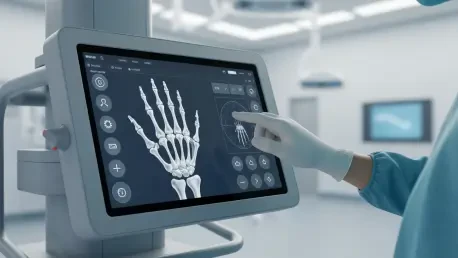
Beyond the core imaging technology, the overall design and user interface of a mini C-arm directly influence both operational efficiency and radiation safety. Features that streamline clinical workflows can significantly shorten procedure times and reduce the need for repeat scans, which in turn contributes to lower cumulative radiation doses for everyone in the room. An intuitive touchscreen interface, for example, simplifies system operation and minimizes the learning curve for staff. Similarly, hands-free control options allow surgeons to manipulate the imaging without breaking the sterile field, enhancing both sterility and speed. Smooth mechanical movement enables quick and precise positioning of the C-arm around the patient, while a compact, ergonomic design facilitates use in crowded surgical suites and reduces physical strain on the clinical team. Efficient digital integration ensures that image capture, storage, and sharing are seamless, minimizing administrative delays and allowing staff to focus more on patient care.
The adoption of imaging platforms designed with radiation awareness in mind yielded significant long-term operational and strategic benefits that redefined the concept of value. Healthcare organizations have moved past assessing equipment based solely on its initial acquisition cost, instead embracing a more holistic, value-based evaluation. The long-term advantages of investing in low-dose mini C-arm technology became clear. These systems provided an enhanced level of safety through a measurable reduction in occupational radiation exposure for surgeons and technicians. They also delivered improved quality and consistency, leading to fewer imaging retakes, which in turn improved both workflow and the patient experience. This technological shift helped facilities meet and exceed increasingly stringent radiation safety standards and aligned with modern value-based care models by prioritizing risk reduction. Ultimately, these advanced systems became the new standard, integrating low-dose protocols without sacrificing the diagnostic clarity essential for excellent patient outcomes.


