
In recent years, remarkable advancements in imaging technologies have dramatically transformed the landscape of cancer treatment and research. These imaging breakthroughs have enabled scientists and clinicians to analyze how cancer cells respond to various drugs simultaneously, marking a leap forward in personalized medicine. By offering tailored and more effective cancer therapies, these innovations herald a new era in oncology focused on precision and individualized treatment protocols. Pioneering this frontier are researchers from the University of Zurich, ETH Zurich, and University Hospital Basel, who are continuously refining these technologies for potential clinical application. The central tenet of their research revolves around sophisticated imaging techniques to visualize and analyze protein behavior within cancer cells. Understanding protein behavior is crucial, as it provides insights into the growth patterns of tumors and their responses to different therapeutic drugs, leading to potentially more effective treatments.
Unraveling Protein Behavior through Advanced Imaging
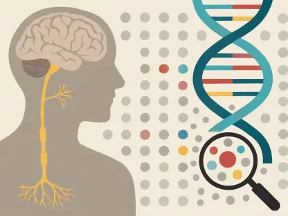
One of the most crucial advancements in imaging technology is the development of fluorescence microscopy techniques, particularly the 4i technology. This innovative method displays the intricate organization of proteins in cancer cells by using dye-labeled molecules that bind specifically to target proteins. Under a fluorescence microscope, scientists can observe where these proteins aggregate within the cells, yielding vital information about the cell’s status. This detailed visualization aids in identifying tumor cell types, offering significant data that inform treatment strategies. Such specificity in identifying and visualizing proteins is critical for formulating targeted cancer therapies, enabling clinicians to tailor treatment plans to the patient’s unique tumor profile. Researchers can also test approximately 50 different cancer drugs simultaneously using these methods. This capability to evaluate drug responses at a molecular level is invaluable for customizing cancer treatments, bringing oncology closer to achieving precision medicine.
Moreover, these imaging techniques pave the way for assessing interactions within cells and their microenvironments, a focus emphasized by Professor Lucas Pelkmans. This understanding can substantially improve the efficacy of cancer treatments by revealing which drug combinations best attenuate growth or induce cell death in tumor cells. The depth of data acquired through advanced microscopy allows for a level of personalization in treatment strategies previously unattainable, offering hope for better therapeutic outcomes.
Progress in Microscopic and Imaging Technologies
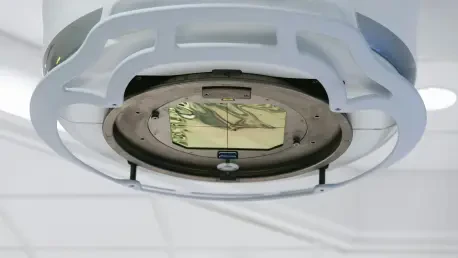
A significant trend in cancer research is the transition from traditional microscopic techniques to sophisticated methodologies like light sheet fluorescence microscopy and imaging mass cytometry. Light sheet fluorescence microscopy has emerged as a powerful tool for observing the gradual development of larger tissue samples over time. This technology supports three-dimensional insights by capturing intricate layers of data, representing an evolutionary leap in bio-imaging. This progression parallels advancements in both hardware and computational methods, offering comprehensive datasets for analysis and interpretation.
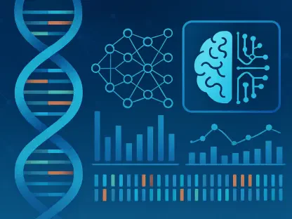
Another crucial development involves artificial intelligence (AI) in processing and analyzing the voluminous data generated by imaging technologies. AI-driven image analysis, or computer vision, facilitates precise interpretations of complex biological images, from cellular size to organelle quantity. Virginie Uhlmann, who leads the BioVisionCenter, highlights the importance of a symbiotic relationship between biological microscopy and computer vision. The development and usage of the Fractal platform exemplify this integration. Fractal allows modular combinations of analytical elements into a seamless workflow, thus facilitating researchers’ access to customized analysis solutions. By democratizing access to advanced image analysis, Fractal enhances research efficiency across the biomedical sphere.
Imaging mass cytometry also plays a pivotal role in contemporary research. Integrating laser and mass spectrometry, this technique offers insights into protein behaviors inside cells. Advanced by Bernd Bodenmiller’s team, it involves labeling antibodies with isotopes to target specific proteins, using a laser to dissect tissue samples for analysis. This method processes samples into a two-dimensional grid, pinpointing protein locales with exceptional accuracy, revealing not only the presence of proteins but elucidating their functional roles within the tumor microenvironment, including immune cell activities.
Transformative Clinical Impacts and Future Directions
Fluorescence microscopy techniques, especially the 4i technology, have revolutionized imaging in the biomedical field. This cutting-edge method utilizes dye-labeled molecules that attach to specific proteins within cancer cells, allowing scientists to see where these proteins collect under a fluorescence microscope. This visualization provides essential insights into the cell’s status and helps in determining the type of tumor cells, delivering valuable information that shapes treatment strategies. The precision in identifying proteins and understanding their organization is instrumental in developing targeted cancer therapies. Such advancements empower clinicians to customize treatments based on an individual’s unique tumor characteristics.
Furthermore, this technology allows researchers to simultaneously test about 50 different cancer drugs. This capability is critical for assessing drug reactions at a molecular level, enhancing the customization of cancer therapies, and advancing oncology towards precision medicine. According to Professor Lucas Pelkmans, these imaging innovations also shed light on the interactions within cells and their surrounding environments. This knowledge significantly improves treatment efficacy by identifying optimal drug combinations to inhibit growth or induce death in tumor cells. The extensive data from advanced microscopy facilitates a level of treatment personalization that once seemed unattainable, providing hope for more successful therapeutic outcomes.