
Imagine a world where scientists can observe the intricate dance of life at the cellular level in real time, capturing every movement of a tiny organism as it navigates its environment, a vision that is no longer a distant dream but a reality thanks to cutting-edge advancements in high-speed 3D microscopy. This technology, exemplified by the groundbreaking M25 microscope, is transforming bioimaging by enabling researchers to study dynamic biological processes with unprecedented detail and speed. The challenge of slow, limited-depth imaging that plagued traditional systems is now being overcome, opening new frontiers in understanding life at its most fundamental level. This review delves into the features, performance, and transformative potential of this remarkable tool in the realm of scientific discovery.
The Evolution of a Game-Changing Technology
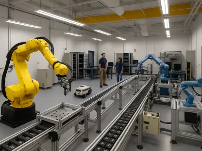
High-speed 3D microscopy has emerged as a pivotal innovation in the field of bioimaging, addressing long-standing limitations of conventional microscopes. Traditional systems often struggled with slow depth scanning and narrow fields of view, missing critical moments in fast-paced biological events. The introduction of high-speed 3D systems marks a paradigm shift, allowing for real-time observation of living organisms in their entirety, capturing dynamic processes as they unfold across multiple dimensions.
At the heart of this evolution lies the need to study life without interruption or distortion. The ability to image entire small organisms, such as worms or fruit flies, in three dimensions without mechanical delays has become essential for fields like developmental biology and neuroscience. This technology integrates advanced optical designs and synchronized imaging to provide a comprehensive view, setting a new standard for how biological research is conducted.
The significance of this advancement extends beyond mere speed. It represents a broader movement in scientific tools toward precision and accessibility, ensuring that researchers can explore complex systems with minimal interference. By enabling non-invasive imaging, high-speed 3D microscopy paves the way for deeper insights into natural behaviors and physiological responses, fundamentally altering the landscape of live specimen studies.
Key Features of the M25 Microscope
Advanced Diffractive Optics for Seamless Imaging
The M25 microscope stands out due to its innovative use of diffractive optical elements, particularly multi-focus gratings, which play a crucial role in its high-speed capabilities. These components manipulate incoming light to create distinct focal planes, allowing simultaneous imaging at various depths without the need for mechanical scanning. This design eliminates delays and distortions commonly associated with traditional systems, ensuring smooth and accurate 3D captures.
Beyond speed, diffractive optics contribute to the compactness and scalability of the M25 system. Unlike bulkier alternatives like prisms, these elements are etched with nanometer-scale precision, making them easier to integrate into existing microscope setups. The result is a streamlined tool that maintains high image quality while pushing the boundaries of what is possible in real-time bioimaging.
This technology also reflects a commitment to precision in optical design. By carefully controlling light distribution, the M25 achieves distortion-free imaging across a wide field of view, a critical factor for studying dynamic processes in small organisms. Such innovation underscores the potential for further refinements in optical engineering to enhance imaging tools.
Multi-Camera Array and Chromatic Precision
Another standout feature of the M25 is its integration of a 25-camera array, designed to capture synchronous images across multiple focal planes. This setup enables the system to record over 100 3D volumes per second within a substantial field of view, providing a holistic perspective of live specimens. The sheer volume of data captured in real time offers researchers an unparalleled ability to analyze rapid biological events.
To maintain clarity at such high speeds, the M25 employs customized blazed gratings for chromatic dispersion correction. This mechanism counters color distortions that can occur when light is split across different focal planes, ensuring consistent resolution and accuracy. The attention to detail in addressing chromatic challenges highlights the system’s dedication to delivering reliable imaging results.
The synergy between the camera array and chromatic correction creates a robust platform for high-speed imaging. This combination not only enhances performance but also sets a benchmark for future microscopy systems aiming to balance speed with precision. Such technical prowess makes the M25 a leading example of innovation in the field.
Performance and Real-World Applications
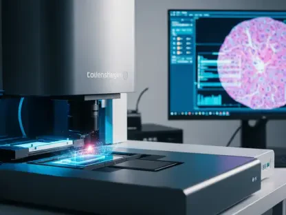
The performance of the M25 microscope is nothing short of revolutionary, particularly in its application to live imaging of small model organisms like C. elegans and D. melanogaster. Capable of observing entire organisms in 3D as they move naturally, the system provides critical insights into neural control of locomotion and developmental processes. This holistic view was previously unattainable with traditional methods that focused on limited sections of a specimen.
Versatility is another hallmark of this technology, as it supports both fluorescence and label-free imaging modalities such as brightfield and polarization microscopy. This compatibility is especially valuable in fields like embryology, where preserving the natural state of delicate specimens is paramount. The ability to conduct minimally invasive studies broadens the scope of research possibilities, from genetic mutation analysis to drug response evaluation.
Moreover, the M25’s design facilitates integration with standard commercial microscopes, requiring no specialized hardware beyond its diffractive optics. With fabrication instructions for optical components publicly available and software for data synchronization accessible on open platforms, the system promotes widespread adoption. This accessibility ensures that labs worldwide can leverage its capabilities to advance their studies, democratizing cutting-edge bioimaging technology.
Challenges in Broader Implementation
Despite its impressive capabilities, high-speed 3D microscopy faces hurdles in scaling to larger organisms or adapting to non-laboratory settings. The current field of view and depth range, while ideal for small model systems, may require significant modifications to accommodate bigger specimens, posing technical challenges in optical design and data processing. Addressing these limitations remains a key focus for ongoing development.
Another obstacle lies in managing the enormous data volumes generated by high-speed imaging. The M25’s ability to capture vast amounts of information in real time demands robust storage and processing solutions, which may strain resources in smaller research facilities. Developing efficient data handling protocols is essential to ensure the technology’s practicality across diverse environments.
Logistical barriers, such as the need for specialized training to operate and maintain advanced systems, also hinder adoption. Efforts are underway to simplify user interfaces and provide comprehensive support resources, aiming to make the technology more approachable for a wider audience. Overcoming these challenges will be crucial for realizing the full potential of high-speed 3D microscopy in varied research contexts.
Looking Ahead: Future Innovations
The future of high-speed 3D microscopy holds immense promise, with potential advancements poised to further revolutionize bioimaging. Integration with machine learning stands out as a key area of exploration, where algorithms could analyze imaging data to identify cell states, track behaviors, or detect disease markers automatically. Such developments would enhance the technology’s utility in biomedical research, streamlining complex analyses.
Additionally, ongoing trends toward scalability and accessibility suggest that future iterations of systems like the M25 could become even more user-friendly and adaptable. Collaborative initiatives to refine optical components and software over the coming years, from now until 2027, aim to lower costs and simplify implementation, ensuring broader reach across academic and industrial sectors. This trajectory reflects a commitment to shared progress in scientific tools.
The long-term impact of these innovations could redefine methodologies for studying biological dynamics. As high-speed 3D microscopy evolves, it may unlock new ways to explore intricate life processes, from cellular interactions to organism-wide responses. Keeping pace with these advancements will be vital for researchers aiming to stay at the forefront of discovery in an ever-changing field.
Final Reflections
Looking back, the journey of high-speed 3D microscopy, as embodied by the M25 system, marked a turning point in how biological processes were visualized and understood. Its ability to capture real-time, three-dimensional dynamics with precision reshaped research in developmental biology and neuroscience. For those eager to harness this technology, the next steps involve exploring open-access resources for fabrication and software, integrating the system into existing lab setups, and collaborating with peers to address scaling challenges. Staying engaged with emerging machine learning applications offers a pathway to maximize data insights, ensuring that the legacy of this innovation continues to inspire and drive scientific progress.