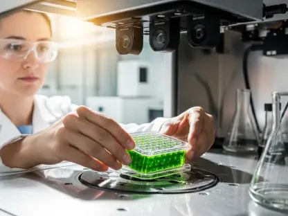

The field of breast MRI has seen remarkable advancements, heralding a new era in breast cancer screening and diagnosis. These innovations not only enhance diagnostic capabilities but also improve the efficiency and comfort of the patient experience. Technological breakthroughs such as abbreviated protocols, AI-assisted analysis, and molecular diagnostics are at the forefront of these developments.
Technological Advances in Breast MRI
Enhanced Role in Detection and Diagnosis
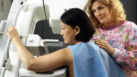
Recent improvements in breast MRI have significantly expanded its diagnostic reach. Radiographer Hanna Kalliomäki from Finland highlighted these advancements at ECR 2025. While mammography remains a staple, MRI is increasingly vital for women with dense breast tissues, affecting nearly half of the screening population. With dense breast tissues, the sensitivity of mammography decreases, often necessitating supplemental imaging techniques to ensure an accurate diagnosis. This growing need has spurred rapid advancements in breast MRI technology, making it an indispensable tool in contemporary breast cancer care.
MRI’s increased usage among women with dense breast tissues is underpinned by its superior ability to differentiate between benign and malignant lesions. Enhanced imaging technology has improved the detection rates of early-stage cancers, facilitating timely and effective treatment. As radiographers continue to refine MRI techniques, they are also tasked with addressing the associated challenges. Ensuring patients are comfortable and properly positioned is crucial to obtaining high-quality images, which directly impacts the success of further analyses, including those involving AI. As breast MRI technology advances, the role of radiographers becomes more critical, demanding continual professional development and adaptation to new methodologies.
Abbreviated Breast MRI (AB-MRI)
A revolutionary development, Abbreviated Breast MRI (AB-MRI), now reduces scan times from up to an hour to just ten minutes. This protocol maintains diagnostic accuracy, increases patient throughput, and decreases anxiety and motion artifacts. Radiographers must adapt to these shorter scans while ensuring they remain precise and informative. The ability to conduct more scans within the same timeframe not only optimizes resource utilization but also allows for broader patient access to advanced imaging. AB-MRI is particularly beneficial for high-risk patients who require frequent monitoring, as it reduces the physical and emotional burden of lengthy scan sessions.
Moreover, the introduction of AB-MRI necessitates adjustments in training and operational workflows for radiographers. They must master the new protocols and understand the nuances of optimizing scan parameters for speed without sacrificing image quality. This requires a deep understanding of the balance between technical efficiency and diagnostic thoroughness. Additionally, patient education becomes paramount, as radiographers play a pivotal role in explaining the benefits and process of AB-MRI to alleviate concerns. As AB-MRI becomes a standard practice, radiographers’ ability to effectively communicate and manage patient expectations will be essential to its successful implementation.
AI-Assisted Analysis
Revolutionizing Image Data Analysis
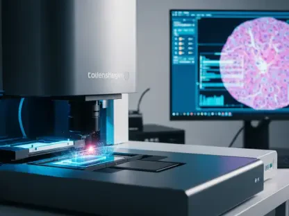
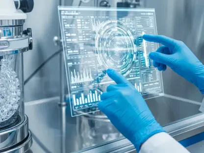
Artificial intelligence (AI) has transformed the way breast MRI images are analyzed. AI tools can identify subtle anomalies, reducing both false positives and false negatives. This technological ally is not meant to replace radiographers but to augment their efficiency and diagnostic precision. The integration of AI into breast MRI promises higher accuracy in detecting early-stage cancers that may be overlooked by human eyes alone. AI algorithms are trained to recognize patterns and deviations in imaging data, offering a second layer of scrutiny that enhances the overall reliability of diagnoses.
However, the effective use of AI in breast MRI requires meticulous quality control and comprehensive training for all involved medical professionals. Radiographers must ensure that patient positioning and image acquisition are performed with impeccable precision, as the quality of the AI interpretation heavily depends on the quality of the initial images. Additionally, radiographers need to stay updated with ongoing advancements in AI technology to leverage its full potential. This includes understanding the operation of AI tools, their limitations, and how to effectively integrate them into routine imaging workflows.
Radiographer Roles and AI Integration
Despite the rise of AI, radiographers remain indispensable. They ensure the quality of scans and correct patient positioning, both critical for effective AI analysis. Understanding the integration of AI into imaging workflows is crucial for radiographers. The collaborative effort between human expertise and AI technology results in a more robust diagnostic process, combining the strengths of both to provide the most comprehensive care. Radiographers’ ability to interpret AI outputs and provide contextually relevant insights based on their clinical experience adds significant value to the diagnostic process.
Moreover, the introduction of AI tools into breast MRI workflows necessitates continuous professional development for radiographers. They must stay abreast of new AI technologies, understand how to calibrate AI systems for optimal performance, and troubleshoot any issues that may arise during the imaging process. Radiographers also need to play an educational role, guiding patients through the new diagnostic landscape and helping them understand the role of AI in their care. By balancing technical proficiency with compassionate patient interaction, radiographers ensure that AI serves as a beneficial complement to their expertise, enhancing overall patient outcomes.
Contrast-Free Imaging and Personalized Profiles
Safer, Contrast-Free Imaging Innovations
Advances like Diffusion-Weighted Imaging (DWI) eliminate the need for contrast agents, making them safer for patients with allergies or kidney issues. DWI highlights abnormal water molecule movements, distinguishing benign from malignant tissues without contrast. This innovation significantly reduces the risk associated with contrast agents, broadening the scope of MRI applicability to a more diverse patient population. By focusing on the natural diffusion of water molecules within tissues, DWI offers a non-invasive, reliable method for identifying pathological changes, particularly in patients who cannot tolerate traditional contrast agents.
Furthermore, the implementation of DWI requires radiographers to adapt their technical skills to optimize imaging settings for different patients. Proper adjustment of DWI parameters is crucial to obtaining high-quality diagnostic images. Radiographers must be adept at troubleshooting and customizing scans based on individual patient needs to maximize the efficacy of this contrast-free technique. This includes understanding the underlying principles of water diffusion in tissues and being able to interpret the resultant images accurately.
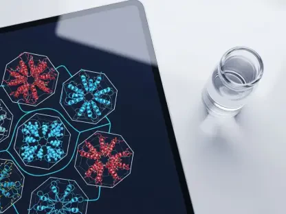
Molecular MRI for Personalized Imaging
Molecular MRI represents another shift toward personalized imaging. Targeted contrast agents that bind to specific cancer cells offer a detailed view of the tumor’s molecular characteristics. This bespoke approach supports targeted therapies, especially for aggressive types like HER2-positive breast cancer. By visualizing tumor-specific markers, molecular MRI facilitates precision medicine, allowing for treatment plans tailored to the unique molecular profile of each patient’s cancer. This advancement not only improves the accuracy of diagnoses but also enhances the effectiveness of subsequent treatments, potentially leading to better patient outcomes.
Radiographers are integral to the successful implementation of molecular MRI. They must be knowledgeable about the various targeted agents used and understand the protocols for their safe administration. This includes being aware of potential side effects and ensuring that the imaging process is as comfortable and safe as possible for patients. Additionally, radiographers must stay informed about the latest developments in molecular imaging to maintain a high standard of care and adapt to new methodologies as they emerge.
Emerging Techniques: Spectroscopy and CDI
Breast MRI Spectroscopy
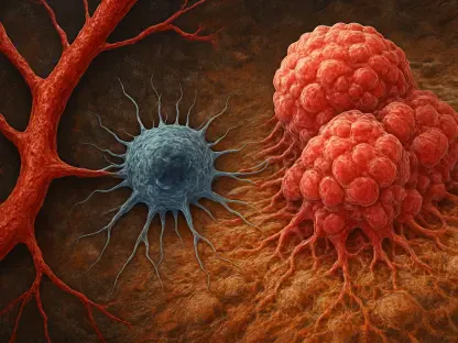
Spectroscopy is an emerging technique analyzing the breast tissue’s chemical composition. It identifies metabolites elevated in tumors, adding another layer to the comprehensive breast MRI assessment. This method provides valuable biochemical insights that are not apparent through traditional imaging techniques. By detecting specific metabolic changes associated with cancerous tissues, spectroscopy can enhance the accuracy of early diagnoses and inform treatment strategies. This technique offers a more detailed understanding of the tumor microenvironment, contributing to the development of more effective, tailored treatment plans.
For radiographers, mastering spectroscopy involves gaining a deep understanding of metabolic imaging principles and learning how to interpret complex spectral data. They must be proficient in the operation and calibration of spectroscopy equipment to ensure accurate and reliable results. Additionally, radiographers play a crucial role in integrating spectroscopy into routine clinical practice, working closely with radiologists and oncologists to provide comprehensive patient care. Their expertise in both the technical and clinical aspects of spectroscopy is essential for leveraging this advanced imaging technique’s full potential.
Synthetic Correlated Diffusion Imaging (CDI)
Synthetic Correlated Diffusion Imaging (CDI) offers biochemical insights, synthesizing multiple DWI images from raw data. This technique provides detailed, nuanced information about breast tissue properties beyond structural imaging alone. CDI enhances the diagnostic capability of traditional DWI by combining multiple diffusion-weighted images into a single, comprehensive dataset. This allows for a more accurate assessment of tissue characteristics, identifying subtle differences that may indicate the presence of malignancies. The synthesis of data from various diffusion imaging perspectives provides a multifaceted view of the breast tissue, aiding in more precise diagnoses.
Radiographers must be skilled in using CDI to maximize its diagnostic benefits. This involves understanding the technical aspects of synthesizing diffusion-weighted images and accurately interpreting the complex data sets produced. Additionally, radiographers need to be adept at adjusting imaging protocols to suit different clinical scenarios, ensuring that CDI is used effectively for each patient. Continuous professional development and staying updated on the latest CDI advancements are crucial for radiographers to maintain high standards of care and fully exploit this technique’s diagnostic potential.
Overarching Trends and Challenges
Moving Towards Efficiency and Patient Comfort
The latest advancements in breast MRI point to increased diagnostic accuracy and efficiency. They promise to transform screening processes, especially for women with dense breast tissue, by integrating AI and adopting contrast-free and personalized imaging techniques. These innovations aim to make breast cancer screening more accessible, accurate, and patient-friendly, addressing the limitations of traditional methods and offering new possibilities for earlier detection and better outcomes. The trend towards efficiency and patient comfort is evident in the development of technologies like AB-MRI and AI-assisted analysis, which streamline imaging workflows and reduce patient burden.
Radiographers play a pivotal role in this transformation by adapting to new technologies and ensuring that they are implemented effectively. Their expertise in patient management, technology operation, and data interpretation is crucial to the success of advanced breast MRI techniques. By focusing on patient comfort and diagnostic accuracy, radiographers help bridge the gap between cutting-edge technology and practical clinical application, ensuring that advancements translate into tangible benefits for patients.
Implementation Challenges
Despite the promising advancements, challenges remain, primarily in MRI availability. Collaborative efforts between radiographers and radiologists are essential to optimizing imaging protocols and improving patient care. The successful integration of new technologies into clinical practice requires coordination and cooperation across various healthcare professionals. Radiographers and radiologists must work together to standardize protocols, ensuring that the latest advancements are utilized to their full potential. This collaborative approach is crucial for overcoming implementation hurdles and realizing the benefits of advanced breast MRI techniques.
Additionally, addressing resource constraints and ensuring equitable access to advanced imaging technologies are significant challenges. As new techniques become available, healthcare systems must prioritize investments in infrastructure and training to support their widespread adoption. This includes increasing the availability of MRI machines, improving training programs for radiographers, and fostering a culture of continuous learning and adaptation. By addressing these challenges, the healthcare community can fully leverage the potential of breast MRI advancements to improve patient outcomes and advance breast cancer care.
Technological Prospects and Future Integration
Potential of Emerging Technologies
Molecular diagnostics and AI-enhanced imaging present a future where cancer detection and treatment become increasingly personalized. Spectroscopy and CDI, although still in their nascent stages, offer promising avenues for more detailed and accurate breast tissue analysis. These technologies provide a more profound understanding of breast cancer at both macroscopic and microscopic levels, leading to earlier detection and more targeted treatments.
Radiographers, by staying at the forefront of these innovations, will play a crucial role in integrating these cutting-edge tools into everyday clinical practice. Continuous professional development and adaptation to new methodologies are essential for maximizing these technologies’ potential.
Ensuring Equity and Accessibility
As these advancements evolve, ensuring equitable access to these technologies will be paramount. Healthcare systems must address potential disparities in availability and training to make these innovations widely accessible. Collaborative efforts between policymakers, healthcare providers, and technology developers will be crucial in overcoming these barriers and ensuring that all patients can benefit from advancements in breast MRI.
By addressing these challenges and fostering collaboration, the future of breast MRI promises to bring significant improvements in diagnostic accuracy, patient comfort, and overall breast cancer outcomes. As the field progresses, the combined efforts of radiographers, radiologists, and other healthcare professionals will be essential in realizing the full potential of these groundbreaking technologies.
Conclusion
The field of breast MRI has experienced significant advancements, ushering in a new era for breast cancer screening and diagnosis. These cutting-edge developments not only improve the ability to diagnose breast cancer but also enhance the efficiency and comfort of the patient experience. Among the most notable technological advancements are abbreviated protocols, which streamline the screening process to save time without compromising accuracy. Additionally, AI-assisted analysis offers advanced interpretation of MRI results, providing radiologists with precise, data-driven insights. This ultimately leads to earlier detection and more personalized treatment plans. Furthermore, molecular diagnostics plays a crucial role in these innovations, enabling a more detailed examination of breast tissue at the molecular level. This deeper understanding aids in identifying the specific types and stages of cancer, leading to more targeted therapy options. These breakthroughs collectively represent a significant leap forward in breast MRI technology, promising better outcomes for patients through enhanced accuracy, efficiency, and overall patient experience.