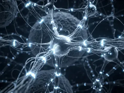
In the fast-evolving field of cellular biology, fluorescence imaging has emerged as a revolutionary tool for tracking cellular processes. With its ability to provide detailed and dynamic views of life on a microscopic scale, it has opened new paths for research and understanding, profoundly changing how scientists study cells. But what makes this technology so significant?
A Brush with History
Fluorescence imaging wasn’t born overnight. The journey began over a century ago when scientists first observed fluorescence—the emission of light by a substance that has absorbed light. However, the leap in technology came with the development of fluorescence microscopy, a groundbreaking technique that uses fluorescent dyes to visualize cellular components.
The significant leap occurred with the introduction of the green fluorescent protein (GFP) from the jellyfish Aequorea victoria. This discovery, which was honored with the Nobel Prize in Chemistry in 2008, allowed scientists to tag proteins within living organisms, providing a way to observe biological processes in real-time.
Major Achievements
Unveiling Cellular Dynamics
The major achievement of fluorescence imaging is its contribution to our understanding of cellular dynamics. By visualizing cell processes in real-time, scientists have been able to study how cells move, divide, and interact. For example, the technology has offered unparalleled insights into the behavior of cancer cells, enhancing the understanding of metastasis and potentially guiding the development of new therapies.
Advancing Disease Research
Fluorescence imaging has been instrumental in disease research. By tagging specific proteins and observing how they interact within the cell, researchers can detect abnormalities that indicate disease. This has proven particularly powerful in neurodegenerative diseases, where understanding protein misfolding and aggregation can lead to early diagnosis and novel treatments.
Personalized Medicine
Another significant achievement of fluorescence imaging is its contribution to the field of personalized medicine. By enabling precise monitoring of cellular responses to different treatments, the technology helps tailor therapies to individual patients. This is particularly promising in cancer treatment, where it can identify the most effective drugs based on the specific characteristics of a patient’s tumor cells.
Unique Traits
High Sensitivity and Specificity
Fluorescence imaging stands out for its high sensitivity and specificity. The ability to use different fluorophores that can bind to specific molecules allows for the detailed study of cellular components and functions, often down to the level of a single molecule.
Real-Time Observation
The capability for real-time observation is another unique trait. This allows scientists to monitor the dynamics of cellular processes as they happen, offering insights that static images cannot provide. The continuous development of live-cell imaging techniques has made tracking these processes even more sophisticated.
Non-Invasiveness
Fluorescence imaging is a non-invasive technique, making it possible to study living cells without adversely affecting their function. This is crucial for accurate and reliable observations of cellular behavior over time.
Current Status and Future Prospects
As of 2023, fluorescence imaging continues to advance, with new techniques and technologies being developed at a rapid pace. Super-resolution microscopy, for instance, pushes the boundaries of resolution beyond the diffraction limit, allowing visualization of structures within cells at the nanoscale.
Advancements in fluorescent probes, such as the development of near-infrared fluorophores, enhance the depth of imaging in living tissues, offering clearer images with less background noise. Further, integrating artificial intelligence and machine learning into image analysis promises to unlock even deeper insights from fluorescence imaging data.
Revisiting Key Points
Fluorescence imaging for tracking cellular processes has dramatically transformed the landscape of cellular biology. Its high sensitivity and specificity, coupled with real-time, non-invasive observation, have made it an indispensable tool. The strides made in unveiling cellular dynamics, advancing disease research, and fostering personalized medicine underscore its significance. The ongoing innovations promise even more breakthroughs, ensuring that fluorescence imaging will remain at the forefront of cellular biology research.
For further resources and up-to-date information, academic journals and professional conferences in the fields of cellular and molecular biology offer extensive content on the latest advancements and applications of fluorescence imaging.


