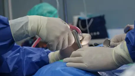
In the ever-evolving landscape of medical technology, a staggering statistic underscores a pressing challenge: gliomas, which constitute 81% of malignant intracranial tumors, often evade complete surgical removal due to the difficulty in distinguishing tumor tissue from healthy brain matter during operations. This critical issue, compounded by brain shifts that occur mid-surgery, frequently results in residual disease and tumor recurrence, posing severe risks to patient outcomes. Among these, glioblastoma, a particularly aggressive subtype, accounts for 45% of gliomas and carries a disheartening five-year survival rate of just 5%. The introduction of a novel imaging solution by an Italian medical imaging company marks a significant step forward in addressing these obstacles. This advancement, showcased at a prominent neurosurgical congress, promises to enhance precision and safety in brain tumor surgeries, potentially transforming the field with cutting-edge technology tailored for real-time intraoperative use.
Revolutionizing Intraoperative Imaging
Addressing Surgical Challenges with Innovation
The complexity of glioma surgeries has long been a hurdle for neurosurgeons, primarily due to the limitations of traditional imaging methods like ultrasound and CT scans. Ultrasound, while non-invasive, suffers from a narrow field of view that can obscure critical details, making it difficult to assess the full extent of a tumor. CT scans, on the other hand, expose patients to repeated doses of ionizing radiation, raising concerns about long-term safety, especially during extended procedures. Closed MRI systems, though more advanced, often come with high costs, operational complexity, and the potential to prolong surgery times, which can elevate risks for patients. These gaps in existing technology highlight a dire need for a solution that offers clarity, safety, and efficiency. The newly introduced open MRI system steps into this void, designed specifically to integrate seamlessly into the surgical environment, offering a fresh approach to overcoming the persistent challenges of distinguishing and removing tumor tissue effectively during operations.
Enhancing Precision Through Real-Time Imaging
A standout feature of this innovative open MRI system is its ability to provide real-time imaging throughout the surgical process, a capability that sets it apart from conventional systems. By incorporating a specialized surgical table and MRI-compatible accessories, the technology allows patients to remain in a single position from start to finish, eliminating the need for repositioning that can introduce errors or delays. This streamlined setup empowers surgeons to confirm the complete removal of tumor tissue before concluding the procedure, significantly reducing the likelihood of residual disease that often necessitates secondary surgeries. Furthermore, the system’s design focuses on minimizing procedural complexity, which not only shortens operation times but also enhances overall patient safety. Such advancements reflect a thoughtful response to the nuanced demands of neurosurgery, where every moment and decision can profoundly impact a patient’s recovery and quality of life, paving the way for more confident and precise surgical interventions.
Transforming Neurosurgical Outcomes
Streamlining Procedures for Better Results
One of the most compelling aspects of this groundbreaking MRI technology is its potential to streamline neurosurgical procedures, thereby improving clinical outcomes for patients facing life-threatening conditions like gliomas. The open design contrasts sharply with the cumbersome nature of traditional closed MRI systems, offering a less intimidating and more accessible setup for surgical teams. This accessibility translates into practical benefits, such as reduced setup times and a lower barrier to adoption in various hospital settings. Clinical experts have noted that the ability to perform imaging at every stage of surgery allows for immediate verification of tumor resection, enabling surgeons to make real-time adjustments with unprecedented accuracy. This iterative process ensures that the maximum amount of tumor tissue is removed while preserving critical healthy brain structures, safeguarding neurological functions. As a result, the technology not only boosts surgical precision but also prioritizes patient well-being in high-stakes environments.
Driving Accessibility and Sustainability in Healthcare
Beyond its technical prowess, this MRI system represents a broader movement toward sustainable and cost-effective solutions in medical technology, addressing the economic challenges often associated with advanced surgical tools. By offering a more user-friendly and less resource-intensive alternative to existing intraoperative imaging systems, the technology stands to democratize access to cutting-edge neurosurgical care, particularly in facilities that may have previously found such equipment prohibitive. The emphasis on sustainability aligns with current trends in healthcare, where balancing clinical effectiveness with practicality is increasingly vital. Insights from neurosurgical leaders highlight how this system integrates seamlessly into daily practice, supporting a vision of innovation that doesn’t compromise on affordability or ease of use. This strategic focus suggests a future where high-quality care becomes more widely available, potentially reshaping how complex surgeries are approached globally and ensuring that more patients benefit from the latest advancements in medical imaging.
Reflecting on a Milestone in Medical Innovation
Looking back, the debut of this open MRI system at a major neurosurgical congress marked a defining moment in the journey toward safer and more effective brain tumor surgeries. The technology addressed longstanding issues with precision and patient safety, setting a new standard for intraoperative imaging that many in the field had long awaited. Moving forward, the focus should shift to broader implementation and continued collaboration between technology developers and clinical practitioners to refine and expand the system’s applications. Exploring how this innovation can be adapted for other complex surgical fields could further amplify its impact. Additionally, ongoing research and feedback from real-world use will be crucial in optimizing its features to meet evolving needs. This milestone serves as a reminder of the power of targeted innovation to solve critical healthcare challenges, offering a blueprint for future advancements that prioritize both technological excellence and tangible improvements in patient care.


