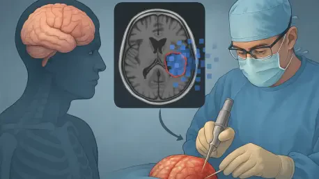

The delicate architecture of the human brain remains remarkably vulnerable to the violent forces of traumatic impact, often triggering a cascading failure of physiological autoregulation that clinicians struggle to contain. Traumatic brain injury (TBI) stands as a premier global health crisis, frequently resulting in permanent neurological deficits or mortality when intracranial pressure escapes medical control. One of the most perilous complications is the development of refractory intracranial hypertension, a condition where the brain swells within the rigid confines of the skull, threatening to crush vital structures. When initial surgical interventions and medical therapies fail to stabilize this pressure, surgeons are forced to perform a secondary decompressive craniectomy. This high-stakes procedure involves removing a substantial portion of the skull to provide the expanding brain tissue with the necessary space to swell without causing a fatal herniation.
Historically, the decision-making process for such a secondary intervention has been almost entirely reactive, relying on visible neurological decline or the crossing of dangerous thresholds on pressure monitors. This traditional “wait and see” strategy creates an incredibly narrow window for surgical action, often leaving medical teams in a desperate race against irreversible tissue damage. However, recent scientific advancements are beginning to dismantle these limitations by leveraging the hidden depth of medical imaging data. Research led by Dr. Zhongyi Sun at Central South University demonstrates that the marriage of radiomics and machine learning can forecast the need for these salvage operations long before clinical symptoms manifest. By identifying the subtle precursors of surgical failure, this technology promises to transform neurotrauma management from a series of emergency reactions into a structured, proactive discipline.
The Science of Radiomics and Machine Learning
Harnessing Hidden Data in Medical Imaging
Radiomics represents a fundamental shift in the utility of medical imaging, moving beyond qualitative visual assessment to treat standard CT scans as vast reservoirs of quantitative data. While a radiologist typically focuses on identifying visible hemorrhages or midline shifts, radiomic analysis utilizes sophisticated algorithms to extract hundreds of features that are invisible to the naked eye. These features include voxel-level descriptors of signal intensity, spatial distribution patterns, and the complex morphology of injured brain tissue. By quantifying these elements, clinicians can objectively measure the microenvironmental heterogeneity of a brain injury. This data provides a granular map of how different regions of the brain are reacting to trauma, allowing for a much more precise estimation of how cerebral edema and hemorrhagic lesions will evolve over the critical first hours of hospitalization.
In the pivotal study conducted by the research team, the focus was narrowed to the primary hemorrhagic lesions and the surrounding edema, which are the primary drivers of dangerous pressure increases. By retrospectively analyzing the pre-surgical imaging of dozens of patients who had undergone an initial craniotomy, the researchers identified specific radiomic signatures that were consistently present in those who eventually required a second surgery. These signatures act as early warning signals, reflecting underlying physiological stress and tissue instability that traditional imaging interpretations often overlook. This level of detail provides a nuanced understanding of the injury’s trajectory, enabling a shift away from subjective assessments toward a standardized, data-driven approach that captures the unique biological fingerprint of every individual’s brain trauma.
Integrating Algorithms for Better Outcomes
The true transformative power of this research is realized through the seamless integration of these high-dimensional radiomic datasets with advanced machine learning algorithms. By feeding complex imaging variables into computational models, researchers can automate the identification of patterns that correlate with specific clinical outcomes, such as the failure of an initial bone-flap replacement. These algorithms are capable of sifting through hundreds of variables simultaneously, recognizing subtle interdependencies between tissue texture and lesion shape that would be impossible for a human brain to process in real-time. This computational bridge effectively translates raw, abstract data into actionable clinical insights, providing a reliable tool that can support neurosurgeons during the intense, high-pressure decision-making environments common in trauma centers.
When these high-tech models were rigorously compared against traditional clinical indicators, the superiority of the radiomic approach became strikingly evident. Standard markers like the Glasgow Coma Scale (GCS) or pupil reactivity, while useful for initial triage, frequently fail to reflect the internal physiological shifts that lead to sudden intracranial pressure spikes. The study found that the most accurate predictive results were generated by a synergistic model that merged radiomic features with specific clinical data points. This hybrid approach offers a holistic view of the patient’s condition, combining the “hidden” signals from the brain tissue with the “visible” symptoms of the patient’s neurological state. This synergy ensures that the predictive model remains grounded in clinical reality while benefiting from the immense analytical depth provided by modern artificial intelligence.
Impact on Clinical Practice and Healthcare Systems
Transitioning to a Proactive Care Model
The ability to accurately predict the necessity of a secondary surgery marks a monumental transition from reactive emergency medicine to a proactive neurosurgical paradigm. If a clinical team can identify a high-risk patient 24 to 48 hours before an intracranial pressure crisis occurs, the entire management strategy undergoes a fundamental reorganization. Instead of waiting for the patient to deteriorate, physicians can initiate aggressive pharmacological interventions, such as tailored osmotic therapy, or adjust ventilator settings to manage carbon dioxide levels more precisely. In many cases, this foresight allows for the secondary decompressive craniectomy to be scheduled as a planned, controlled procedure during daylight hours. This avoids the inherent risks of emergency salvage operations performed under suboptimal conditions when the patient is already in a state of physiological collapse.
By intervening early in the progression of the injury, medical teams can effectively mitigate the severity of secondary brain injury. This refers to the secondary cascade of chemical, metabolic, and physical damage that follows the initial impact, often causing more long-term harm than the trauma itself. A proactive approach not only preserves more viable brain tissue but also significantly enhances the chances of the patient regaining functional independence and cognitive clarity. Shifting the management focus in this manner ensures that neurosurgeons remain ahead of the pathology, transforming the “golden hour” of trauma into a “golden window” of sustained, data-informed care. This evolution fundamentally changes the survival narrative for TBI patients, moving the goalpost from mere survival to the preservation of a high quality of post-traumatic life.
Resource Management and Systemic Benefits
Implementing predictive radiomics models also yields significant advantages for the broader healthcare infrastructure, which is often strained by the immense costs of neurotrauma care. Traumatic brain injuries are among the most resource-intensive conditions to treat, requiring extended stays in the intensive care unit (ICU), continuous invasive monitoring, and multiple rounds of surgery. By using radiomic scores to identify patients at low risk for complications, hospital administrators and department heads can allocate expensive resources more efficiently. Critical equipment, such as intracranial pressure monitors, and highly sought-after ICU beds can be prioritized for those whom the data suggests are most likely to face life-threatening swelling. This targeted allocation ensures that the most vulnerable patients receive the highest level of care without unnecessary expenditures on those who are stable.
Furthermore, reducing the frequency of emergency re-operations has a stabilizing effect on the entire hospital ecosystem. Emergency surgeries often disrupt elective schedules, place extreme stress on surgical staff, and increase the likelihood of medical errors due to fatigue and urgency. A more predictable surgical workflow leads to better staff retention and improved overall safety protocols within the operating theater. On a socio-economic scale, improving the neurological outcomes for TBI survivors—who are frequently young individuals in the prime of their working lives—reduces the long-term burden on family caregivers and state-funded social welfare programs. By utilizing technology to optimize the path to recovery, healthcare systems can alleviate the profound financial and emotional weight that traumatic injuries place on society as a whole.
Future Horizons in Precision Neurosurgery
Automated Pipelines and Artificial Intelligence
The trajectory of neurosurgery is clearly leaning toward a data-driven future where artificial intelligence serves as a central pillar of critical care. The ultimate objective for researchers like Dr. Sun is the creation of fully automated diagnostic pipelines that integrate directly into hospital imaging software. In such a system, the moment a CT scan is finalized, an AI algorithm would immediately process the images, extract the relevant radiomic features, and generate a standardized risk score. Within minutes, the surgical team would receive an alert regarding the patient’s probability of requiring secondary intervention. This provides a digital “second opinion” backed by thousands of previous cases, helping to eliminate the variability in care that often exists between different trauma centers or shifts of medical personnel.
This type of automated safety net would be especially transformative for regional or smaller trauma centers that may lack 24-hour access to specialized neurosurgical expertise. By providing a reliable, algorithmic flag for high-risk cases, AI-driven radiomics ensures that critical warning signs are never overlooked regardless of the attending physician’s experience level. This vision brings the concept of “time is brain” to a new level of clinical reality, using advanced technology to ensure that every second is used to defend the patient’s neurological integrity. As these systems become more sophisticated, they will likely incorporate real-time data from other bedside monitors, creating a comprehensive, living model of the patient’s physiological state that can alert doctors to trouble before it even starts to surface on a monitor.
Overcoming Challenges and Validating the Path
While the initial results of radiomic research are undeniably promising, several significant hurdles must be cleared before this technology becomes a global standard of care. Currently, many of these predictive models have been developed using data from single institutions, which raises questions about their generalizability across different patient populations and ethnicities. To address this, the next phase of development must involve large-scale, multi-center validation trials to ensure that the algorithms remain accurate regardless of where they are deployed. Furthermore, there is a technical need for cross-platform compatibility, as the software must function seamlessly across various CT scanner manufacturers and different hospital picture archiving and communication systems (PACS) without requiring extensive manual data entry.
Refining these algorithms is an ongoing process that requires constant calibration to ensure they are robust enough for diverse clinical scenarios. As researchers continue to validate these tools through 2027 and 2028, the movement toward “precision neurosurgery” will gain irreversible momentum. This approach ensures that no two patients are treated exactly the same, as every treatment plan will be uniquely tailored to the specific quantitative profile of their injury. By replacing generalized protocols with individualized, data-driven strategies, the medical community can offer a new sense of hope to those facing the devastating reality of brain trauma. This path forward represents the pinnacle of modern medicine: the use of human ingenuity and computational power to preserve the dignity, personality, and future of those whose lives have been suddenly altered by injury.
In the years following the initial trauma, the primary goal of medical intervention was often limited to simple survival, yet the integration of radiomics and machine learning has shifted that focus toward functional restoration. This research demonstrated that the hidden patterns within a simple CT scan could hold the key to preventing the most catastrophic outcomes of brain swelling. By providing surgeons with the foresight to act before a crisis occurred, these tools effectively reduced the incidence of emergency re-operations and improved the overall recovery rates for patients. The transition to a proactive management model proved to be a vital step in modernizing neurotrauma care, ensuring that clinical decisions were based on objective data rather than reactive observations. Ultimately, these advancements established a new standard for precision neurosurgery, where technology and human expertise worked in tandem to protect the most complex organ in the human body.


