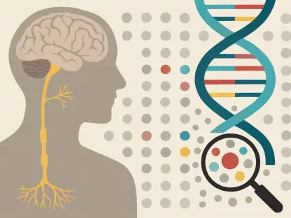
Ivan Kairatov is a leading biopharma expert specializing in the intersection of biotechnology and clinical innovation. With extensive experience in research and development, he has spent his career bridging the gap between laboratory breakthroughs and practical healthcare solutions. In this discussion, we explore the revolutionary potential of cf-EpiTracing, a platform developed by researchers at PKU that promises to redefine non-invasive diagnostics. We delve into how epigenetic fingerprints can pinpoint the origins of disease from a single drop of blood, the role of machine learning in ensuring diagnostic accuracy, and the future of multi-omic tools in monitoring patient health with unprecedented precision.
Clinical tests often require large blood volumes, but it is now possible to diagnose conditions using only 50 microliters of plasma. How does this impact testing for pediatric or elderly patients, and what technical steps ensure that a single drop provides enough data for a reliable result?
The ability to work with just 50 microliters of plasma—roughly a single drop of blood—is a monumental shift for vulnerable populations like infants and the elderly, where traditional large-volume blood draws can be physically taxing or technically difficult. For a premature infant in the NICU, every milliliter of blood is precious, and for elderly patients with fragile veins, this minimally invasive approach reduces both physical trauma and procedural anxiety. The technical reliability of the cf-EpiTracing platform hinges on its ability to capture high-density epigenetic signals that were previously lost during standard processing. By focusing on the rich information contained within cell-free chromatin rather than just fragmented DNA, the system amplifies the “signal-to-noise” ratio, ensuring that even a tiny sample provides a comprehensive biological snapshot. This shift means we no longer need quantity to achieve quality; we just need the right molecular lens to view the sample.
Identifying the exact tissue where a disease originates remains a major hurdle for non-invasive screenings. How do epigenetic fingerprints help pinpoint these cellular origins, and what are the practical advantages of using cell-free chromatin over traditional DNA-based liquid biopsies?
Traditional liquid biopsies often focus on DNA mutations, which tell us that a disease exists but frequently fail to reveal exactly where in the body it is located. Epigenetic fingerprints act like a cellular return address, revealing the specific tissue of origin because different cells leave unique marks on their chromatin when they enter the bloodstream. By analyzing cell-free chromatin, we can see the regulatory state of the cells that shed that material, allowing us to trace a signal directly back to the colon, the lungs, or the bone marrow. This provides a much more nuanced view than simple DNA sequencing, as it captures the functional identity of the tissue. Practically, this allows clinicians to move beyond a “positive” or “negative” result and instead pinpoint the specific site of concern, which is essential for targeted follow-up imaging or biopsies.
Screening for colorectal cancer has recently reached accuracy rates as high as 97% in controlled settings. How do machine learning algorithms maintain this performance when moving to independent validation groups, and what specific metrics determine if these models are ready for widespread clinical use?
Maintaining high performance outside of a controlled training environment is the ultimate test for any diagnostic AI, and it was impressive to see this platform hold a robust 92.2% accuracy in independent validation groups after achieving 97.6% in the training phase. These machine learning algorithms succeed by integrating multimodal epigenomic features, which prevents the model from “overfitting” or becoming too focused on the quirks of a single small data set. We look for consistency across different patient demographics and stages of disease to ensure the tool is truly generalizable. The primary metrics for clinical readiness are sensitivity and specificity—essentially, how well the tool avoids missing a cancer case while also avoiding the emotional and financial cost of false positives. When a model maintains over 90% accuracy across diverse, independent cohorts, it signals that the underlying biological signals are stable and reliable enough for the real world.
In cases of diffuse large B cell lymphoma, specific plasma signals can indicate bone marrow involvement and disease aggressiveness. How does detecting CD34-positive cells in blood change subtyping strategies, and what are the implications for designing personalized treatment plans for these patients?
The discovery that patients with diffuse large B cell lymphoma exhibit stronger signals of CD34-positive cells in their plasma is a potential game-changer for how we categorize this aggressive cancer. CD34 is a marker typically associated with hematopoietic stem cells, and its presence in the blood suggests that the lymphoma may have already begun to involve the bone marrow, signaling a more advanced or aggressive disease state. This allows oncologists to subtype the disease more accurately without always needing invasive bone marrow biopsies, which are notoriously painful for the patient. By identifying these high-risk signals early through a simple blood draw, we can design personalized treatment plans that might include more intensive therapies or earlier interventions. It moves us away from a “one-size-fits-all” treatment for lymphoma and toward a strategy tailored to the specific cellular dynamics of the individual’s cancer.
Integrating DNA methylation and chromatin topology into a single platform creates a sophisticated multi-omic diagnostic tool. What are the logistical trade-offs when combining these complex data layers, and how will this approach transform the way clinicians monitor cellular changes during a patient’s treatment?
Combining DNA methylation, mutations, and chromatin topology into a multi-omic platform offers incredible precision, but it does come with the logistical challenge of managing massive amounts of complex data. Each layer of information requires specialized processing, which can increase the time and cost of the analysis compared to a single-modality test. However, the trade-off is well worth it because this integrated view allows clinicians to monitor the “evolution” of a disease in real-time as a patient undergoes treatment. Instead of waiting months for a tumor to shrink on a scan, we can observe shifts in the cellular dynamics and epigenetic markers in the blood to see if a therapy is working within weeks. This transformative approach turns the blood into a continuous live-stream of the patient’s internal health, allowing for rapid adjustments to treatment plans that could ultimately save lives.
What is your forecast for the future of non-invasive disease tracing?
I believe we are entering an era where the traditional “yearly physical” will be replaced by deep-dive molecular profiling from a single drop of blood. Within the next decade, I forecast that platforms like cf-EpiTracing will become the gold standard not just for cancer, but for detecting neurodegenerative and cardiovascular diseases years before physical symptoms appear. We will see a shift toward proactive, rather than reactive, medicine, where the “epigenetic clock” of various organs is monitored to catch cellular dysfunction at its absolute infancy. This will democratize high-end diagnostics, making sophisticated disease tracing accessible in primary care clinics and even remote areas, fundamentally changing our relationship with our own biological data.