
In a world where early detection of health conditions can mean the difference between life and death, a groundbreaking development in medical imaging technology is capturing the attention of researchers and clinicians alike. Emerging from the labs at the University of Colorado Boulder, a novel non-mechanical optical coherence tomography (OCT) device is poised to transform how eye and heart diseases are diagnosed. This innovative system, which replaces traditional moving parts with electrowetting liquid lenses, offers a glimpse into a future where high-resolution imaging is more reliable, compact, and accessible. By steering light through voltage adjustments instead of physical components, the technology not only enhances durability but also significantly reduces power consumption. As healthcare continues to prioritize early intervention, this advancement could bridge critical gaps in diagnostics, potentially saving countless lives through timely and accurate detection of conditions that often go unnoticed until it’s too late.
Breaking Down the Technological Innovation
Redefining OCT with Non-Mechanical Design
A significant leap forward in medical imaging, the new OCT system developed at the University of Colorado Boulder addresses longstanding challenges associated with traditional setups. Conventional OCT devices, widely used for detailed imaging of biological tissues, rely on mechanical components like spinning mirrors to direct light waves, making them prone to wear and tear while demanding substantial power. The introduction of electrowetting liquid lenses changes this dynamic entirely by using voltage to steer light, eliminating the need for moving parts. This shift results in a more robust and energy-efficient design, capable of withstanding the rigors of frequent use without the risk of mechanical failure. Such an approach not only extends the lifespan of the equipment but also paves the way for smaller, more portable units that could be deployed in diverse medical settings, from specialized clinics to remote areas lacking advanced infrastructure.
Advantages of Miniaturization and Efficiency
Beyond durability, the non-mechanical design of this OCT system unlocks remarkable potential for miniaturization, a critical factor in expanding its applications. The elimination of bulky components allows the technology to be integrated into ultra-thin catheters, flexible endoscopes, and even wearable devices, making it adaptable for various diagnostic needs. This compactness is complemented by reduced power requirements, which enhance the feasibility of long-term or portable use, particularly in scenarios where energy resources are limited. For patients, this translates to less invasive procedures and greater comfort, as smaller tools can navigate the body with minimal disruption. Additionally, the efficiency of the system supports continuous health monitoring, a growing trend in personalized medicine. By enabling real-time imaging in compact formats, this technology could redefine how medical professionals approach diagnostics, bringing precision closer to the point of care than ever before.
Expanding Applications in Healthcare
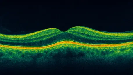
Transforming Eye Care with Enhanced Imaging
The implications of this non-mechanical OCT technology for ophthalmology are profound, promising earlier and more accurate detection of retinal disorders. Conditions such as macular degeneration and diabetic retinopathy, which can lead to irreversible vision loss if not caught early, stand to benefit immensely from the high-resolution imaging capabilities of this system. During rigorous testing, the prototype successfully captured subcellular-resolution images of zebrafish eyes, a model often used due to its similarities to human ocular anatomy. These images matched the clarity of commercial OCT systems, resolving intricate structures like the cornea and iris with exceptional precision. Such accuracy could enable clinicians to spot subtle changes in retinal tissue long before symptoms manifest, allowing for interventions that preserve sight. This advancement underscores a potential shift in eye care, where diagnostic tools become more reliable and accessible across various healthcare environments.
Pioneering Cardiac Diagnostics and Beyond
Equally compelling is the potential of this technology to revolutionize cardiovascular diagnostics, an area where early detection is often a matter of life and death. The compact and flexible nature of the non-mechanical OCT system makes it suitable for imaging coronary arteries to identify early signs of atherosclerosis, a leading cause of heart attacks and strokes. By integrating into ultra-thin catheters, the device could provide real-time, high-resolution visuals during minimally invasive procedures, reducing patient discomfort and improving procedural outcomes. Lead researcher Samuel Gilinsky has highlighted the promise of in vivo imaging inside the body, which could transform how cardiologists approach diagnosis and treatment. Furthermore, the technology’s adaptability suggests applications beyond cardiology, potentially extending into neurology and other fields where detailed imaging is critical. This versatility positions the innovation as a cornerstone for future medical advancements, broadening the horizons of non-invasive diagnostics.
Future Pathways and Clinical Translation
Looking ahead, the journey from prototype to clinical application holds immense promise, supported by funding from esteemed organizations like the National Institutes of Health and the National Science Foundation. The research team is already charting a roadmap that includes developing specialized prototypes for retinal and cardiac imaging, aiming to replace the bulkier systems currently in use with streamlined, flexible alternatives. Co-author Juliet Gopinath has emphasized the opportunity to detect health conditions at earlier stages, ultimately improving patient outcomes through timely intervention. While challenges such as regulatory approval and widespread adoption remain, the successful validation of the system’s precision and stability offers a strong foundation for optimism. As the medical field continues to trend toward less invasive and patient-friendly solutions, this technology aligns perfectly with industry priorities, potentially setting a new standard for diagnostic tools in the coming years.
Reflecting on a Diagnostic Milestone
Reflecting on this remarkable development, it becomes evident that the non-mechanical OCT system marks a turning point in medical imaging history. Its ability to deliver high-resolution visuals with enhanced reliability and compactness addresses critical pain points of traditional systems. Testing on zebrafish eyes demonstrated a level of precision that rivaled existing commercial devices, while plans for clinical prototypes signaled a clear intent to impact real-world healthcare. The innovation opens doors to broader applications, from strengthening ophthalmology to pioneering advancements in cardiac care, aligning seamlessly with the push for minimally invasive tools. As a testament to its potential, the technology promises to bring diagnostics closer to patients, whether in clinics or through wearable solutions. Moving forward, the focus should shift to navigating regulatory landscapes and fostering partnerships to ensure this breakthrough reaches those who need it most, setting a precedent for future healthcare innovations.


