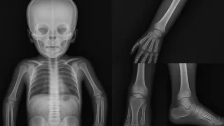

In the intricate world of pediatric radiology, one of the most pressing challenges is determining whether certain bone features in children, specifically in the metaphyseal regions of the distal femurs and proximal tibias, represent normal growth variations or indicate serious pathological lesions. These areas, located near the ends of long bones, are critical zones of development in young patients, and their appearance on imaging can often be misleading. A normal variation might resemble a lesion caused by trauma, metabolic disease, or even malignancy, leading to potential misdiagnosis. This confusion can have profound consequences, ranging from unnecessary stress and medical interventions to delayed treatment of genuine conditions. Recent research has delved into this complex issue, aiming to refine diagnostic accuracy by better understanding the nuances of bone morphology in children. The ability to distinguish between harmless variations and concerning lesions is not just a technical challenge but a vital step toward improving health outcomes for young patients facing skeletal uncertainties.
Unraveling the Complexity of Bone Development
Distinguishing between normal metaphyseal variations and pathological lesions in children’s bones is a task fraught with difficulty due to the dynamic nature of skeletal growth. The metaphyseal regions of the distal femur and proximal tibia are particularly prone to variability as they play a central role in bone lengthening during childhood. Factors such as age, sex, and individual developmental patterns contribute to a wide range of appearances on imaging studies, which can closely mimic signs of serious conditions like fractures or metabolic disorders. Studies underscore the importance of recognizing these normal differences to prevent diagnostic errors. Without a clear framework for identifying what falls within the spectrum of typical growth, radiologists risk misinterpreting harmless features as alarming abnormalities, potentially subjecting young patients to undue stress and medical scrutiny. This highlights a critical gap in current practices that recent research seeks to address through detailed analysis of bone morphology.
The consequences of misdiagnosis in this context extend far beyond mere technical errors, impacting both clinical care and emotional well-being. When a normal variation is mistaken for a lesion, it can trigger a cascade of unnecessary tests, invasive procedures, and heightened anxiety for families already navigating the complexities of pediatric healthcare. Conversely, failing to identify a true lesion may delay essential treatment, allowing underlying conditions to progress unchecked. This dual risk underscores the urgent need for improved diagnostic criteria that can reliably separate typical growth patterns from pathological changes. Research emphasizes that achieving this clarity is not just about enhancing imaging interpretation but also about safeguarding patient trust in medical systems. By focusing on the subtle distinctions in metaphyseal appearance, the field of pediatric radiology aims to minimize errors and ensure that interventions are both timely and appropriate for each child’s unique situation.
Harnessing Technology for Diagnostic Precision
Advancements in imaging technology have emerged as powerful allies in the effort to differentiate metaphyseal variations from lesions, offering unprecedented detail in the analysis of bone structure. High-resolution scans and quantitative assessment tools enable radiologists to examine the intricacies of the distal femur and proximal tibia with greater accuracy than ever before. These innovations reveal subtle textural and structural differences that can help distinguish a benign variation from a concerning abnormality, reducing the likelihood of misinterpretation. Research highlights that integrating such cutting-edge tools into routine practice is essential for elevating diagnostic standards in pediatric radiology. The enhanced visibility provided by modern imaging not only aids in identifying normal growth patterns but also builds confidence among clinicians tasked with making critical decisions. As technology continues to evolve, its role in refining the diagnostic process becomes increasingly indispensable for ensuring young patients receive the most accurate assessments possible.
Beyond the capabilities of advanced imaging, the development of age-specific diagnostic guidelines represents a crucial step toward addressing the variability inherent in children’s bone growth. Skeletal development does not follow a uniform timeline, meaning that what appears atypical in one age group might be entirely expected in another. Tailored references that account for these developmental stages provide radiologists with a contextual framework for interpreting imaging results, ensuring that findings are evaluated against appropriate benchmarks. Studies suggest that such guidelines can significantly reduce diagnostic ambiguity by aligning assessments with the natural progression of bone maturation. This approach acknowledges the diversity of growth patterns among children and emphasizes the importance of customization in medical diagnostics. By adopting age-specific standards, the field moves closer to a model of care that prioritizes precision and minimizes the risk of over- or under-diagnosis in pediatric skeletal evaluations.
Shaping the Future of Pediatric Radiology
The implications of distinguishing metaphyseal variations from lesions extend into the realm of medical education, where equipping future clinicians with specialized knowledge is paramount. Training programs must evolve to include comprehensive instruction on the nuances of bone morphology, particularly in the context of pediatric growth. By fostering an understanding of how normal variations present on imaging and differ from pathological conditions, educational initiatives can help reduce diagnostic errors over the long term. Research advocates for the integration of these insights into curricula, ensuring that emerging radiologists and physicians are well-prepared to navigate the complexities of skeletal assessments. This focus on education aligns with broader trends in healthcare that prioritize continuous learning and skill enhancement, ultimately contributing to a workforce capable of delivering high-quality care. Strengthening the foundation of medical training in this area promises to enhance patient outcomes through informed and accurate clinical decision-making.
Equally significant is the connection between this diagnostic challenge and the principles of personalized medicine, which emphasize tailoring healthcare to individual characteristics. Recognizing that each child’s bone development follows a unique trajectory allows for diagnostic approaches that are customized to specific needs rather than relying on generalized assumptions. This research supports the shift toward precision in radiology by advocating for methods that account for personal variations in skeletal growth. Such an approach ensures that diagnoses are not only accurate but also relevant to the patient’s distinct physiological profile, minimizing the risk of inappropriate interventions. The push for personalization reflects a growing consensus in medicine that effective care must adapt to the individual, particularly in fields like pediatric radiology where developmental differences are pronounced. By embracing this mindset, the discipline can better serve young patients with assessments that are both scientifically sound and deeply considerate of their unique circumstances.
Looking ahead, the exploration of metaphyseal variations opens up numerous avenues for future research that could further transform pediatric skeletal diagnostics. Investigating the roles of genetic and environmental factors in shaping bone morphology offers the potential to uncover deeper insights into why variations occur and how they manifest over time. Longitudinal studies tracking these changes across childhood and adolescence are also critical for mapping the evolution of metaphyseal features, providing data that can refine diagnostic criteria. Research suggests that such investigations could lead to breakthroughs in understanding skeletal health, paving the way for more targeted approaches to managing bone-related conditions. As the field continues to probe these unanswered questions, the promise of enhanced diagnostic tools and strategies looms large, offering hope for even greater accuracy in distinguishing normal growth from pathology. This ongoing commitment to discovery ensures that pediatric radiology remains at the forefront of medical innovation, poised to address emerging challenges with rigor and foresight.
Reflecting on Progress and Next Steps
Reflecting on the strides made in pediatric radiology, it becomes clear that significant progress has been achieved in tackling the challenge of distinguishing metaphyseal variations from lesions. Groundbreaking research has shed light on the importance of recognizing normal growth patterns in the distal femurs and proximal tibias, preventing countless misdiagnoses that could have led to unnecessary interventions. The adoption of advanced imaging technologies has played a pivotal role in enhancing clarity, while age-specific guidelines have provided a vital framework for contextual interpretation. Educational efforts have also laid the groundwork for a more skilled generation of clinicians. Moving forward, the focus should shift to expanding research into genetic and environmental influences on bone development, alongside sustained investment in longitudinal studies. These next steps promise to deepen the understanding of skeletal morphology, ensuring that diagnostic practices evolve in tandem with scientific discovery. By prioritizing innovation and precision, the field stands ready to further improve care for young patients facing skeletal uncertainties.


