
The shift toward precision medicine requires a continuous assessment of risk that updates dynamically as new follow-up scans and blood tests become available. This ongoing demand for agility is driving a fundamental transformation in lung cancer management, moving the field away from isolated clinical evaluations and toward a unified, data-driven framework. For too long, the oncology community has relied on static snapshots of disease—a single CT scan or a solitary biopsy—that often fail to capture the full, evolving complexity of a tumor’s biological profile. These traditional workflows are frequently hindered by the inability to synthesize multidimensional data manually, leading to missed opportunities for early intervention or reactive treatment plans that address symptoms rather than underlying drivers. By leveraging machine learning to convert massive volumes of information from diverse medical sources into actionable evidence, researchers are now creating a more cohesive patient journey. This shift ensures that every clinical decision is backed by comprehensive, quantitative insights rather than fragmented observations. As these technologies mature, the goal is to bridge the gap between various diagnostic tools, providing a holistic view of the disease that was previously unattainable through conventional methods alone. This integration allows for a more nuanced understanding of how malignancy develops and persists over time, transforming the standard of care from a reactive model into a proactive, predictive science that prioritizes individual patient needs through rigorous data analysis.
Synthesizing Multi-Dimensional Clinical Data Streams
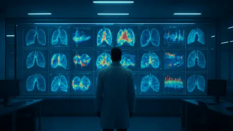
The foundation of modern oncology lies in the integration of five primary data pillars: medical imaging, multi-omics, liquid biopsies, digital pathology, and electronic health records. Radiographic data from CT and PET scans provide the essential spatial context, while molecular profiling through genomics and proteomics uncovers the biological drivers of tumor growth. Meanwhile, liquid biopsies offer a minimally invasive way to monitor disease changes in real-time through blood samples, and digital pathology allows for an unprecedented level of detail in analyzing cellular environments. When these biological and radiological findings are grounded in a patient’s longitudinal history—such as smoking status, environmental exposures, and prior treatment outcomes found in electronic health records—the result is a powerful diagnostic engine. Machine learning acts as the essential connective tissue in this ecosystem, identifying subtle patterns and radiomic features that are nearly invisible to the human eye. This multi-source synergy allows clinicians to move beyond traditional assessments, providing a more robust basis for identifying malignancy and predicting how a specific cancer might behave over time. By centralizing these disparate streams, the medical community can finally address the heterogeneity of lung cancer, recognizing that two tumors appearing identical on a scan may actually require vastly different therapeutic approaches based on their genetic signatures.
Building upon this integrated data environment, the concept of radiomics has emerged as a cornerstone of data-driven lung cancer care. Radiomics involves the high-throughput extraction of quantitative features from medical images, transforming standard scans into mineable data. These features, ranging from tumor texture and shape to the intensity of the surrounding tissue, can reveal information about the tumor microenvironment that traditional visual inspection simply cannot grasp. For instance, a machine learning model might identify a specific “signature” of spiculated margins or internal density variations that correlates strongly with an aggressive mutation, such as an EGFR or ALK rearrangement. By combining these radiomic signatures with the molecular insights gained from liquid biopsies, which track circulating tumor DNA in the bloodstream, physicians can monitor a patient’s response to therapy without the need for repeated, invasive tissue biopsies. This real-time feedback loop is particularly vital in 2026, where the rapid evolution of cancer cells necessitates a constant recalibration of treatment strategies. The synthesis of these diverse data types not only improves the accuracy of the initial diagnosis but also provides a dynamic roadmap for the entire treatment process, ensuring that the clinical team is always one step ahead of the disease’s progression.
Architectural Advancements: From Statistics to Foundation Models
The methodology behind cancer research has evolved from traditional statistical models to sophisticated deep learning architectures that can handle the sheer scale of modern medical data. While early methods like random forests or support vector machines remain useful for smaller, well-defined datasets, they often require human experts to manually select and define which variables to analyze—a process known as feature engineering. In contrast, modern deep learning allows systems to learn directly from raw data, such as high-resolution pathology slides or complex volumetric images. This shift has paved the way for multimodal fusion, where models merge different data types into a single latent space to reach a more accurate clinical conclusion. In this environment, the model does not just look at a CT scan and a genetic report separately; it understands the relationship between a specific imaging pattern and a specific genomic mutation. This allows for a deeper level of inference, where the AI can predict survival outcomes or recurrence risks by identifying cross-modal correlations that would be impossible for a human to synthesize. This architectural shift is fundamental to handling the complexities of lung cancer, where the interplay between the host immune system and the tumor cells is constantly changing.
The latest frontier in this technological evolution is the emergence of foundation models, which are massive AI systems trained on vast, diverse datasets that can be fine-tuned for specific oncology tasks. These models represent a departure from narrow AI, which was typically designed for a single purpose, such as segmenting a lung nodule. Instead, foundation models are “pre-trained” on millions of medical images and text records, allowing them to develop a broad understanding of medical concepts before they are ever applied to a specific patient case. This allows for greater efficiency in clinical settings, as a pre-trained model requires much less specific data to become proficient at a new task, such as identifying rare sub-types of small cell lung cancer. However, as these models become more integrated into healthcare systems, they must overcome the significant hurdle of generalizability. Because medical equipment, imaging protocols, and patient demographics vary across institutions, a model trained in one metropolitan hospital might struggle when deployed in a rural clinic with different technology. Consequently, current research is heavily focused on external validation and transparent reporting, ensuring that these powerful architectures remain reliable across diverse populations and clinical environments, thereby democratizing access to high-tier diagnostic intelligence.
Precision Diagnostics: Transforming Early Detection and Prognosis
Integrating machine learning into the clinical pathway significantly improves early detection, which remains the most critical factor in improving lung cancer survival rates. By analyzing subtle radiological patterns and combining them with liquid biopsy signals, AI systems can more accurately distinguish between benign nodules and early-stage cancer. This precision is essential because traditional screening programs often suffer from high false-positive rates, leading to unnecessary anxiety, cost, and invasive follow-up procedures like lung resections for non-cancerous growths. In 2026, the application of deep learning models to low-dose CT scans has allowed for the identification of high-risk patients long before clinical symptoms appear. These models can detect minute changes in nodule volume or density over time, providing a quantitative measure of growth that exceeds the capabilities of manual measurement. This high-sensitivity approach effectively shifts the medical response from reactive treatment of advanced disease to proactive intervention at a stage when the cancer is most curable. Furthermore, by identifying patients who are at the highest risk based on a combination of imaging and genomic data, healthcare systems can better allocate resources, ensuring that those who need urgent care receive it without delay.
Beyond initial diagnosis, data-driven models enable a level of precision medicine that was previously unattainable through standard clinical guidelines. Because lung cancer consists of biologically distinct subtypes with varying levels of aggressiveness, patients often respond differently to various therapies, such as targeted inhibitors or immunotherapies. AI can match a patient’s unique molecular signature to the most effective drug or surgical option by analyzing historical data from thousands of similar cases. Furthermore, instead of relying on static survival estimates, multi-source data allows for dynamic prognosis. This continuous assessment updates as new follow-up scans or blood tests become available, allowing doctors to detect treatment resistance early. For instance, if an AI model identifies a rise in circulating tumor DNA alongside subtle changes in the texture of a shrinking tumor, it may signal that the cancer is developing resistance to the current drug. This early warning system allows clinicians to adjust strategies, perhaps switching to a secondary treatment line, before the disease progresses significantly. This shift toward a more fluid, data-informed prognosis ensures that treatment remains personalized and effective throughout the entire course of the patient’s care.
Navigating the Path to Clinical Adoption
Despite the clear benefits, several systemic hurdles must be cleared before AI becomes a standard, everyday tool in every oncology clinic across the country. Data standardization remains a primary challenge, as different hospitals and diagnostic centers often use varying protocols for imaging and documentation. This lack of uniformity makes it difficult for machine learning models to function universally, as a model might be “confused” by variations in scan thickness or the specific software used to record pathology results. Additionally, the sheer computational power required to process billions of pixels from high-resolution digital pathology slides can lead to technical complications like overfitting. Overfitting occurs when a model performs exceptionally well on the specific data it was trained on but fails to generalize when faced with real-world, messy data from a different source. To combat this, researchers are focusing on developing more robust data-cleansing techniques and universal standards for medical data reporting. Ensuring that data is “AI-ready” is now a top priority for healthcare administrators who recognize that the quality of the insights provided by an AI system is only as good as the quality of the data fed into it.
Clinician trust is another vital component of adoption, requiring a move toward interpretable AI to solve the notorious “black box” problem. Doctors must understand the underlying reasoning behind an AI’s recommendation to feel confident in using it to guide life-altering patient care decisions. If a model flags a nodule as high-risk, the clinician needs to know if that decision was based on the nodule’s shape, its density, or its relationship to surrounding blood vessels. Developing “explainable” interfaces that highlight the specific regions of an image or the specific genetic markers that influenced the model’s output is essential for building this rapport. Finally, protecting patient privacy is paramount in an era where data is the most valuable resource. Researchers are exploring decentralized solutions like federated learning, where AI models are trained locally within the secure servers of individual hospitals. In this setup, the model “learns” from the data at each site and then shares its learned weights—rather than the raw, sensitive patient records—with a central system. This allows the global AI system to gain knowledge from a wide range of diverse data without ever requiring sensitive information to leave its original, secure location, maintaining the highest standards of patient confidentiality.
Strategic Initiatives for a Data-Driven Oncology Framework
The progress established by late 2026 showed that the primary obstacle to AI integration was not a lack of technology, but a lack of infrastructure. Stakeholders recognized that for these tools to move beyond the research phase, healthcare systems had to prioritize the development of high-speed data pipelines and centralized biobanks. These initiatives facilitated the secure sharing of de-identified patient information, which allowed for the creation of more diverse and representative training sets. By addressing the data silos that previously isolated information within individual departments, organizations enabled a more collaborative approach to cancer research. This infrastructure also supported the deployment of real-time monitoring tools, ensuring that the insights generated by AI models were delivered directly to the clinician’s workstation at the point of care. The focus shifted from simply developing better algorithms to creating an ecosystem where those algorithms could be used reliably and ethically. This systemic overhaul ensured that the power of machine learning was harnessed to its full potential, moving the needle on survival rates by making sophisticated diagnostic tools available to a broader range of clinicians and their patients.
Moving forward, the successful implementation of these systems required a commitment to continuous education and ethical oversight. Medical schools and residency programs began incorporating data science and AI literacy into their curricula, ensuring that the next generation of oncologists would be prepared to work alongside digital assistants. Simultaneously, regulatory bodies updated their frameworks to ensure that AI-driven devices underwent rigorous longitudinal monitoring even after they entered the market. This ensured that any “drift” in a model’s performance was caught and corrected early, maintaining the safety and efficacy of the technology. To further enhance the impact of these tools, future efforts should focus on integrating social determinants of health—such as air quality and socioeconomic factors—into the existing clinical models. By expanding the data pool to include environmental and lifestyle variables, researchers can develop an even more comprehensive understanding of lung cancer risk. This holistic approach, combined with the technical foundations laid in the mid-2020s, will continue to refine the precision of cancer care, ensuring that the transition toward a data-driven oncology framework remains patient-centered and evidence-based for years to come.