
The formidable challenge of treating pancreatic cancer has long been defined by a frustrating cycle of trial and error, where potent chemotherapies are administered with profoundly unpredictable results for individual patients. This clinical uncertainty is a heavy burden in the fight against a disease known for its aggressive nature and grim prognosis. For the vast majority of patients diagnosed with unresectable tumors, chemotherapy is the primary line of defense, yet the selection of the most effective drug regimen has remained more of an educated guess than a precise science. A groundbreaking development, however, now offers the potential to replace this uncertainty with data-driven prediction, leveraging a standard diagnostic scan to foresee how a patient’s unique tumor will respond to a specific treatment, heralding a new era of personalized oncology.
This leap forward addresses one of the most pressing needs in the management of Pancreatic Ductal Adenocarcinoma (PDAC). With a five-year survival rate hovering at a stark 13%, the imperative to optimize every aspect of treatment is paramount. The current standard of care forces oncologists and patients into a difficult gamble, choosing between powerful chemotherapy cocktails like gemcitabine/nab-paclitaxel (AG) or FOLFIRINOX without a reliable method to predict which will be effective. This not only risks wasting precious time with an ineffective therapy but also subjects patients to significant toxic side effects without the corresponding benefit. At the heart of this therapeutic puzzle lies the tumor’s microenvironment, a complex landscape that can either facilitate or thwart treatment, and a new noninvasive tool finally promises to map this crucial terrain.
The Critical Need for a Predictive Breakthrough
Dubbed the “king of cancers” for its lethality and resilience, Pancreatic Ductal Adenocarcinoma stands as one of modern medicine’s most daunting challenges. The disease is notoriously difficult to detect in its early stages and progresses rapidly, leaving many patients with advanced, inoperable tumors at the time of diagnosis. For these individuals, systemic chemotherapy is not just a treatment option but often the only one, making its effectiveness a matter of life and death. The stark reality is that even with the best available therapies, the overall prognosis remains poor, underscoring the urgent need for innovations that can improve outcomes.
This urgency is magnified by the inherent variability in patient response to standard chemotherapy regimens. Two patients with seemingly similar diagnoses can have dramatically different outcomes when given the same drugs. This inconsistency forces clinicians into a reactive, trial-and-error approach where a treatment’s failure is only known after weeks or months of administration, a period during which the disease can progress unchecked. The absence of reliable biomarkers to guide these critical first-line treatment decisions represents a significant gap in patient care, leaving a clear and compelling need for tools that can prospectively identify the right drug for the right patient.
A key reason for this therapeutic variability lies within the unique biology of pancreatic tumors. A defining characteristic of PDAC is extensive fibrosis, the formation of a dense, scar-like connective tissue known as a desmoplastic stroma. This fibrotic matrix is not merely a passive scaffold; it actively participates in tumor growth, metastasis, and immunosuppression. Moreover, it creates a formidable physical barrier, compressing blood vessels and impeding the delivery of chemotherapeutic agents to the cancer cells they are meant to destroy. Understanding and quantifying this fibrosis has long been a goal for researchers, as it holds the key to unlocking more effective treatment strategies.
Establishing Ground Truth with Digital Pathology
Before a noninvasive tool could be developed, researchers first had to establish an objective and highly accurate method for measuring tumor fibrosis, creating a “ground truth” against which any new technology could be benchmarked. Traditional methods relying on biopsies are inherently limited. An invasive biopsy provides only a small tissue sample, which may not accurately represent the heterogeneity of the entire tumor, leading to significant sampling errors. This limitation has historically hindered the development of fibrosis as a reliable clinical biomarker.
To overcome this challenge, a multicenter team of researchers turned to the power of artificial intelligence and digital pathology. They analyzed a vast collection of whole-slide tumor images from 361 patients who had undergone surgery for PDAC. Using sophisticated deep learning algorithms, they trained a computer model to precisely segment the tumor tissue and objectively quantify the proportion of fibrotic stroma within it. This automated process eliminated the subjectivity of manual assessment and provided a reproducible, quantitative score for fibrosis for each tumor in its entirety.
The initial findings from this digital pathology work were profoundly important. Across several independent patient cohorts, this AI-quantified fibrosis level proved to be a powerful prognostic biomarker. Intriguingly, the data revealed that patients with highly fibrotic tumors tended to have significantly longer overall survival. This discovery, confirmed through transcriptome analysis that linked high fibrosis to pathways involved in collagen metabolism, validated that an objective measure of the stromal environment provides critical clinical information, setting a solid foundation for the next stage of the research.
Translating Pathology into a Noninvasive Scan
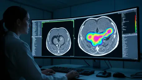
With a validated “ground truth” for fibrosis established, the next crucial step was to translate this complex pathological insight into a simple, noninvasive clinical tool. The goal was to develop a method that could measure fibrosis without requiring a surgical tissue sample. For this, the research team leveraged a diagnostic tool that is already a routine part of the clinical workflow for nearly every pancreatic cancer patient: the preoperative contrast-enhanced computed tomography (CT) scan. This approach ensures that the resulting model can be easily integrated into standard practice without adding procedures, costs, or delays.
The innovative technique centered on the field of radiomics, which involves extracting vast amounts of quantitative data from medical images that are invisible to the naked eye. The researchers extracted thousands of these radiomic features from the preoperative CT scans, capturing subtle variations in tumor texture, shape, intensity, and other complex patterns. This deep data mining allows the computer to “see” underlying biological characteristics that are not apparent to a human radiologist reviewing the same scan.
From this immense pool of data, the team employed advanced machine learning techniques to identify the 15 most critical radiomic features that were most strongly correlated with the actual fibrosis levels measured by the digital pathology gold standard. These 15 features were then combined to build a predictive model capable of generating a CT-based fibrosis score from a standard scan. The model’s performance was rigorously tested and validated on an external cohort of patients, where it achieved an area under the curve (AUC) of 0.718, indicating a high degree of accuracy and reliability in predicting tumor fibrosis noninvasively.
A Landmark Finding for a Common Chemotherapy
The true clinical power of this new CT-based model was revealed when it was applied to a cohort of 295 patients with unresectable pancreatic cancer who were undergoing first-line chemotherapy. The study, published in the journal Research, aimed to determine if the noninvasively predicted fibrosis score could do more than just predict prognosis; the goal was to see if it could predict a patient’s response to a specific treatment, transforming it from a prognostic tool into a predictive one.
The results were dramatic and unequivocal. For patients treated with the gemcitabine/nab-paclitaxel (AG) chemotherapy regimen, the CT-predicted fibrosis level was directly linked to treatment efficacy. Patients whose scans indicated high-fibrosis tumors experienced a near-doubling of their median overall survival, which soared from 7.73 months to an impressive 13.37 months. Their median progression-free survival also saw a significant increase, extending from 4.70 months to 6.23 months. This demonstrated that patients with a highly fibrotic tumor microenvironment derive a profound and clinically meaningful benefit from AG therapy.
Critically, this powerful correlation was exclusive to the AG regimen. When the researchers analyzed patients treated with other common chemotherapies, such as FOLFIRINOX or SOXIRI, they found no significant link between the CT fibrosis score and treatment outcomes. This specificity is what elevates the finding from a simple observation to a landmark discovery. It provides, for the first time, a noninvasive biomarker that can specifically identify the patient population most likely to respond to gemcitabine/nab-paclitaxel, offering a clear, data-driven rationale for personalizing treatment decisions in a disease where every choice matters.
Charting a Path From the Lab to the Clinic
The most compelling aspect of this research is its immediate potential for clinical translation. Because the predictive model relies solely on standard preoperative CT scans, it can be implemented without altering existing diagnostic protocols. The technology is designed for seamless integration into hospital Picture Archiving and Communication Systems (PACS), the software platforms that radiologists and oncologists already use daily. This would place a powerful decision-support tool directly into the hands of clinicians, allowing them to rapidly assess a patient’s tumor fibrosis profile and make more informed choices about first-line chemotherapy.
Beyond guiding the selection of existing therapies, these insights into the tumor microenvironment can also inform the development of novel and more effective treatment strategies. For instance, in patients with high-fibrosis tumors who are already primed for a superior response to AG, it may be possible to further enhance that response by combining the chemotherapy with matrix-targeting agents. These emerging drugs are designed to break down the dense stromal barrier, potentially improving drug penetration and making the cancer cells even more vulnerable to treatment.
The methodological framework established by this study provides a powerful blueprint for the future of precision oncology. The model’s accuracy could be further refined by incorporating data from other imaging modalities like MRI or PET scans and by leveraging next-generation AI algorithms. Furthermore, the core concept of noninvasively quantifying the tumor microenvironment is not limited to pancreatic cancer. This approach holds immense promise for other fibrotic cancers, including breast, colorectal, and ovarian cancers, potentially creating a new paradigm for predicting treatment response across a wide spectrum of malignancies.
This pioneering work successfully demonstrated how artificial intelligence could decode the complex biological information hidden within a routine medical scan. By translating a key feature of the tumor microenvironment into a clinically actionable biomarker, the research has provided a tangible tool that could reshape the therapeutic landscape for one of the world’s deadliest cancers. The findings represented a critical step away from a one-size-fits-all approach and toward a future where treatment is tailored to the unique biology of each patient’s disease, offering renewed hope for better outcomes.


