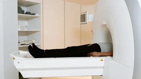
The utilization of artificial intelligence (AI) in medical imaging is revolutionizing the way diseases are detected and diagnosed, providing groundbreaking opportunities to identify conditions beyond the primary purpose of the scans. A pioneering study led by researchers at NYU Langone Health has demonstrated the incredible potential of AI tools to identify heart disease by using computed tomography (CT) scans that were initially taken for other medical reasons. This innovative approach, known as “opportunistic screening,” involves repurposing existing medical images to uncover additional health issues that were not the primary focus of the original scan.
Leading the way in this new frontier, the study showcased at the Radiological Society of North America (RSNA) meeting highlighted how AI could measure the amount of aortic calcium in routine abdominal CT scans. By assigning a standard score to the calcification level, the AI tool could predict an individual’s risk of experiencing major cardiovascular events such as heart attacks or vessel blockages. Dedicated CT scans of coronary arteries have traditionally been used to identify heart diseases. However, these types of scans are infrequent and often not covered by insurance, making the AI-enhanced analysis of routine abdominal scans a significant move forward in early detection and preventive care for heart disease.
AI Detects Heart Disease in Routine CT Scans
Researchers evaluated 3,662 CT scans taken between 2013 and 2023 from older individuals in the New York area. These participants had both abdominal scans and dedicated coronary artery CT scans. The findings indicated that the AI-enabled analysis of calcification in the aorta could accurately predict the extent of calcification in the coronary arteries and the associated risk of major cardiovascular events. This study suggests that abdominal scans alone could serve as a valuable predictive tool for heart attacks and other cardiovascular issues, allowing for earlier and more frequent detection.
The study’s results demonstrated that individuals with aortic artery calcification were 2.2 times more likely to experience significant cardiovascular events such as heart attacks, brain vessel blockages, or the need for procedures to restore heart blood flow over a three-year monitoring period. Out of the study’s participants, 324 individuals encountered such events. Moreover, early signs of arterial calcium buildup were detected in 29 percent of participants who were previously thought to have none, underscoring the potential of this AI-assisted screening in identifying at-risk individuals much earlier than traditional methods allow.
Early Detection and Preventive Care
This new finding corroborates earlier research published in the journal Bone, which also explored the use of opportunistic screening. In this prior study, Dr. Miriam A. Bredella and her team examined CT scans intended for lung cancer screening and discovered significant bone loss in patients, pointing out the underdiagnosis of osteoporosis. The underdiagnosis was notably higher among racial minority groups. The study revealed that osteoporosis was present in 38 percent of Black patients, 55 percent of Asian patients, 56 percent of Hispanic patients, and 72 percent of White patients screened, bringing to light significant disparities in the detection and diagnosis of the disease.
The opportunistic screening tool in the previous study also identified high ratios of body fat, arterial hardening, and fatty liver, which are factors associated with bone loss. This additional insight emphasizes the potential for AI tools to diagnose multiple conditions using a single scan, which could be particularly valuable for vulnerable or underserved populations who often lack access to specialized screenings. These findings suggest a wider application of AI tools in routine scans could play an essential role in early disease detection and intervention, providing a new frontier in preventive health care.
Broader Applications of Opportunistic Screening
Dr. Bredella emphasized that the research supports the use of opportunistic screening as an effective method for diagnosing and treating conditions like osteoporosis and heart disease, particularly in populations at higher risk, such as the elderly and smokers. This innovative approach has the potential to bridge gaps in access to preventive care and improve health outcomes by enabling earlier intervention. By repurposing routine medical images to identify additional health issues, opportunistic screening can uncover conditions that might otherwise go unnoticed until they become more severe and harder to treat.
However, while the findings are promising, Dr. Bredella noted the necessity for further research to confirm whether the imaging data and analyses are sufficient for the early identification and effective treatment of major coronary diseases and osteoporosis. Such validation is vital for ensuring that this technology can reliably reduce illness and mortality rates. Ensuring the accuracy and reliability of AI-assisted screenings will also be crucial for their broader application in clinical practice, providing healthcare professionals with the tools they need to deliver better patient care.
Need for Further Research and Validation
The use of artificial intelligence (AI) in medical imaging is transforming how diseases are detected and diagnosed, offering groundbreaking opportunities to identify conditions beyond the primary purpose of the scans. A cutting-edge study led by researchers at NYU Langone Health has shown AI’s immense potential in identifying heart disease through computed tomography (CT) scans initially taken for other medical reasons. This technique, known as “opportunistic screening,” repurposes existing medical images to uncover additional health issues not originally targeted by the scan.
At the Radiological Society of North America (RSNA) meeting, the study demonstrated how AI could measure aortic calcium levels in routine abdominal CT scans. The AI tool assigns a standard score to the calcification, enabling it to predict an individual’s risk of major cardiovascular events like heart attacks or vessel blockages. Traditional scans of coronary arteries are typically used for heart disease detection, but they are rare and often not covered by insurance. Thus, AI-enhanced analysis of routine abdominal scans marks a significant advance in early detection and preventive care for heart disease.


