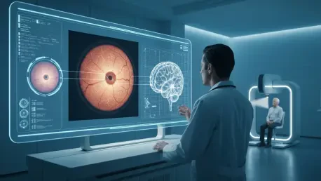

A groundbreaking new methodology that harnesses the power of artificial intelligence to analyze retinal fundus images is poised to revolutionize how medical professionals assess brain health, offering a non-invasive, cost-effective, and rapid alternative to traditional neuroimaging techniques. This innovative approach is built upon the well-established biological principle that the retina, with its intricate network of blood vessels and neural tissue, serves as an accessible window to the brain. By examining the back of the eye, clinicians can detect the earliest signs of cognitive decline and other neurological conditions, potentially transforming a routine eye exam into a vital screening tool for some of the most challenging diseases of our time. This paradigm shift promises to move brain health monitoring from specialized, expensive procedures like MRI or CT scans into a more accessible, preventative framework, making early detection a reality for a much broader population.
The Science Behind the Scan
The Retina-Brain Connection
The profound anatomical and physiological link between the eye and the brain forms the scientific bedrock of this innovative diagnostic approach. As a direct extension of the central nervous system, the retina contains a dense network of neural tissue and blood vessels that share developmental origins and structural characteristics with the brain. This intimate connection means that pathological changes occurring within the brain, particularly those associated with neurovascular or neurodegenerative conditions, can often manifest as subtle, yet detectable, alterations in the retina’s delicate microvasculature and nerve layers. This bio-physiological mirroring positions the retina as a uniquely reliable proxy for cerebral health, allowing for the observation of systemic changes without requiring invasive procedures. The clinical significance of this connection is now widely accepted, with a growing consensus that the eye can provide invaluable insights into neurological well-being, transforming ophthalmology’s role in comprehensive patient care.
This direct biological relationship establishes retinal fundus imaging not merely as a convenient alternative but as a potentially superior tool for certain diagnostic purposes when compared to conventional methods. The ability to visualize and quantify changes in retinal blood vessels or the optic nerve provides a direct line of sight into the health of the body’s neurovascular system. For example, the narrowing of retinal arterioles or the presence of micro-aneurysms can mirror similar processes happening in the brain’s less accessible blood vessels, offering early warnings for conditions like stroke or vascular dementia. Similarly, the thinning of the retinal nerve fiber layer has been strongly correlated with neurodegenerative diseases such as Alzheimer’s and Parkinson’s. This non-invasive visibility into neural and vascular health offers a powerful advantage, providing clinicians with actionable data that can guide preventative strategies and early interventions, long before more severe and irreversible symptoms emerge in patients.
A Leap Beyond Traditional Methods
When compared directly with conventional brain health diagnostics, the advantages of retinal imaging become starkly clear. Established methods like Magnetic Resonance Imaging (MRI) and Computed Tomography (CT) scans, while powerful, present numerous logistical and financial hurdles. These procedures are often expensive, placing a significant burden on both healthcare systems and patients. They also require substantial time commitments for the scan itself and for the subsequent analysis by a radiologist. Furthermore, access can be limited by the availability of specialized equipment and trained personnel, creating disparities in care, particularly in rural or underserved communities. For certain patient populations, such as those with claustrophobia or metallic implants, these scans can be inaccessible or highly distressing. In sharp contrast, retinal fundus imaging is a highly efficient, cost-effective, and rapid assessment tool. The procedure is quick, requires no special patient preparation, and can be performed with relatively portable equipment, making it an ideal candidate for widespread screening and routine monitoring programs.
The practical benefits of integrating retinal imaging into standard health assessments could fundamentally reshape the landscape of neurological care. Its accessibility and affordability make it suitable for large-scale screening initiatives that can identify at-risk individuals long before they would typically seek specialist consultation. This shifts the paradigm from reactive diagnosis, which often occurs after significant cognitive decline, to a proactive model focused on prevention and early intervention. For healthcare systems, this means a potential reduction in the long-term costs associated with managing advanced neurodegenerative diseases. For patients, it offers the promise of earlier diagnosis, which is critical for treatments that are most effective in the initial stages of a disease. This technology could empower primary care physicians and optometrists on the front lines of healthcare, equipping them with a powerful tool to monitor the neurological health of their patients as part of a routine check-up, thereby democratizing access to brain health evaluation.
The Power of Intelligent Imaging
Fusing AI with Clinical Insight
This diagnostic breakthrough is not solely a product of the retina-brain connection but is profoundly enabled by the powerful convergence of two critical technological elements: advanced imaging hardware and sophisticated artificial intelligence. Recent progress in optical engineering has dramatically increased the clarity and resolution of retinal scans, allowing for the detection of subtle biomarkers that were previously imperceptible to the human eye. This high-fidelity data, capturing minute details of the retinal landscape, provides the raw material for deep analysis. This is where AI becomes indispensable. Complex algorithms, trained on vast datasets of retinal images correlated with known neurological outcomes, can identify intricate patterns, anomalies, and pathological changes with a level of accuracy and objectivity that often surpasses human capabilities. This synergy between capturing better data and applying smarter analysis unlocks the full potential of the retina as a diagnostic window.
A pivotal innovation that elevates this technology from a simple screening tool to a sophisticated diagnostic aid is the integration of “clinical information prompts.” This methodology enriches the AI’s analysis by providing crucial context, transforming the assessment from a generic evaluation into a highly personalized one. Instead of analyzing an image in isolation, the system leverages specific patient data—such as medical history, presenting symptoms, genetic predispositions, and other known risk factors—to guide and refine its diagnostic process. This prompt-driven approach allows the AI to weigh certain visual features more heavily based on a patient’s individual profile, leading to a more nuanced and accurate interpretation. This not only aids in confirming diagnoses for existing conditions but, more importantly, facilitates the early identification of at-risk individuals who could benefit from preventative measures and timely intervention, making it a powerful tool for personalized medicine.
Revolutionizing Geriatric Care
The societal implications of this advanced diagnostic technology are particularly profound in the context of a globally aging population. The rising incidence of neurodegenerative diseases, including Alzheimer’s and Parkinson’s, has created an urgent and growing demand for diagnostic tools capable of early and accurate detection. The proposed method directly addresses this critical need. By enabling the prompt identification of cognitive decline, often before severe and debilitating symptoms manifest, it opens a crucial window for interventions that can be most effective in slowing disease progression and improving a patient’s long-term quality of life. This capability is invaluable for geriatric healthcare providers, as it offers a practical, accessible means to screen for neurological risk as part of a standard health assessment for older adults. The technology has the potential to become a cornerstone of preventative geriatric medicine, fundamentally changing how cognitive health is monitored and managed in later life.
Beyond individual patient benefits, the widespread adoption of this AI-powered retinal screening could have a transformative impact on the entire healthcare ecosystem. By providing a low-cost, high-throughput method for assessing brain health, it could alleviate the significant strain currently placed on specialized neurology departments and expensive imaging facilities. Primary care physicians and optometrists could be empowered to conduct initial screenings, allowing for more efficient triage of patients and ensuring that specialist resources are directed toward those who need them most. This decentralization of diagnostic capability could lead to more equitable access to care, reduce wait times for specialist appointments, and ultimately lower the overall economic burden of neurodegenerative diseases. By integrating this technology into routine care, healthcare systems can build a more resilient and proactive infrastructure for managing the neurological health challenges of an aging world.
Charting the Course for the Future
Validation and New Research Horizons
To substantiate their claims and build a solid foundation for clinical adoption, the researchers behind this method conducted an extensive analysis using a robust dataset sourced from a diverse cohort of participants. This rigorous, empirical approach enabled them to establish strong, statistically significant correlations between specific retinal alterations and key neurological markers, solidifying the scientific argument for this technique. The success of this research also serves as a powerful testament to the indispensable role of interdisciplinary collaboration. By combining the deep expertise of ophthalmologists, who understand the eye; neurologists, who understand the brain; and data scientists, who build the AI models, the project created a synergy that led to a true breakthrough. This collaborative model is presented as a blueprint for future medical innovation, demonstrating how integrating diverse knowledge bases is essential for solving complex health challenges.
Beyond its immediate and promising clinical applications, this study has opened several exciting new avenues for future research that could further deepen our understanding of the brain. A more granular investigation into the specific biological pathways and molecular mechanisms that connect retinal pathology with cerebral dysfunction could uncover entirely new therapeutic targets for neurodegenerative disorders. This could pave the way for the development of novel treatments designed to interrupt disease processes at a much earlier stage. Furthermore, future studies could explore a compelling and potentially transformative question: could interventions designed to improve or protect retinal health also confer a therapeutic benefit for cognitive function? If this bi-directional link is confirmed, it could lead to a dual-impact approach to brain health, where protecting vision also means protecting the mind.
Navigating Ethical Challenges
While the technological promise of AI-driven diagnostics is immense, its integration into healthcare must be navigated with a responsible acknowledgment of the inherent ethical considerations. Critical issues such as the security and privacy of sensitive patient data are paramount. Robust frameworks must be established to ensure that this information is protected from misuse or breaches. Moreover, the need for algorithmic transparency—often referred to as the “black box” problem—must be addressed. Clinicians and patients alike need to have confidence in and a basic understanding of how the AI reaches its conclusions to foster trust and ensure accountability. Finally, the potential for inherent biases in machine learning models, which are trained on existing datasets that may reflect historical health disparities, must be rigorously identified and mitigated to ensure that these tools promote equitable access to care rather than exacerbating existing inequalities.
Ultimately, the research into prompt-driven retinal imaging laid a robust foundation for a significant evolution in medical diagnostics. The integration of this technology into routine neurological assessments represented a paradigm shift, moving healthcare away from siloed specializations and toward more integrated, comprehensive, and patient-centered models of care. By embracing this innovative potential, healthcare systems significantly enhanced their ability to monitor, maintain, and promote neurological health on a global scale. This work did not just introduce a new tool; it fundamentally changed the future of brain health evaluation by making it more accessible, proactive, and personalized. The path forward was paved with the promise of earlier diagnoses and better outcomes for millions of individuals worldwide.


